File:Gray1024.jpg
Original file (800 × 691 pixels, file size: 117 KB, MIME type: image/jpeg)
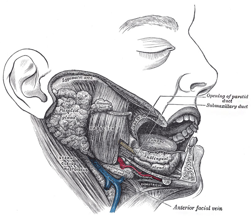
Fig. 1024. Dissection showing Salivary Glands of right side
Three large pairs of salivary glands communicate with the mouth, and pour their secretion into its cavity; they are the parotid, submaxillary, and sublingual.
parotid gland (glandula parotis) (Figs. 1022, 1023), the largest of the three, varies in weight from 14 to 28 gm. It lies upon the side of the face, immediately below and in front of the external ear. The main portion of the gland is superficial, somewhat flattened and quadrilateral in form, and is placed between the ramus of the mandible in front and the mastoid process and Sternocleidomastoideus behind, overlapping, however, both boundaries. Above, it is broad and reaches nearly to the zygomatic arch; below, it tapers somewhat to about the level of a line joining the tip of the mastoid process to the angle of the mandible. The remainder of the gland is irregularly wedge-shaped, and extends deeply inward toward the pharyngeal wall.
submandibular gland (or Submaxillary Gland (glandula submaxillaris)) is irregular in form and about the size of a walnut. A considerable part of it is situated in the submaxillary triangle, reaching forward to the anterior belly of the Digastricus and backward to the stylomandibular ligament, which intervenes between it and the parotid gland. Above, it extends under cover of the body of the mandible; below, it usually overlaps the intermediate tendon of the Digastricus and the insertion of the Stylohyoideus, while from its deep surface a tongue-like deep process extends forward above the Mylohyoideus muscle.
sublingual gland (glandula sublingualis) is the smallest of the three glands. It is situated beneath the mucous membrane of the floor of the mouth, at the side of the frenulum linguæ, in contact with the sublingual depression on the inner surface of the mandible, close to the symphysis. It is narrow, flattened, shaped somewhat like an almond, and weighs nearly 2 gm. It is in relation, above, with the mucous membrane; below, with the Mylohyoideus; behind, with the deep part of the submaxillary gland; laterally, with the mandible; and medially, with the Genioglossus, from which it is separated by the lingual nerve and the submaxillary duct. Its excretory ducts are from eight to twenty in number. Of the smaller sublingual ducts (ducts of Rivinus), some join the submaxillary duct; others open separately into the mouth, on the elevated crest of mucous membrane (plica sublingualis), caused by the projection of the gland, on either side of the frenulum linguæ. One or more join to form the larger sublingual duct (duct of Bartholin), which opens into the submaxillary duct.
- Links: Tongue Development | Salivary Gland Development | Head Development | Musculoskeletal System Development |
- Gray's Images: Development | Lymphatic | Neural | Vision | Hearing | Somatosensory | Integumentary | Respiratory | Gastrointestinal | Urogenital | Endocrine | Surface Anatomy | iBook | Historic Disclaimer
| Historic Disclaimer - information about historic embryology pages |
|---|
 |
| iBook - Gray's Embryology | |
|---|---|

|
|
Reference
Gray H. Anatomy of the human body. (1918) Philadelphia: Lea & Febiger.
Cite this page: Hill, M.A. (2024, April 19) Embryology Gray1024.jpg. Retrieved from https://embryology.med.unsw.edu.au/embryology/index.php/File:Gray1024.jpg
- © Dr Mark Hill 2024, UNSW Embryology ISBN: 978 0 7334 2609 4 - UNSW CRICOS Provider Code No. 00098G
File history
Click on a date/time to view the file as it appeared at that time.
| Date/Time | Thumbnail | Dimensions | User | Comment | |
|---|---|---|---|---|---|
| current | 08:34, 11 May 2014 | | 800 × 691 (117 KB) | Z8600021 (talk | contribs) | :'''Links:''' Tongue Development | Salivary Gland Development | Head Development | Musculoskeletal System Development | {{Gray Anatomy}} Category:Tongue Category:Head Category:Cartoon |
You cannot overwrite this file.
File usage
The following 4 pages use this file: