File:Gray1022.jpg
Gray1022.jpg (629 × 400 pixels, file size: 45 KB, MIME type: image/jpeg)
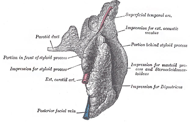
Fig. 1022. Right parotid gland (posterior and deep aspects)
Parotid Gland (glandula parotis) (Figs. 1022, 1023, 1024), the largest of the three, varies in weight from 14 to 28 gm. It lies upon the side of the face, immediately below and in front of the external ear. The main portion of the gland is superficial, somewhat flattened and quadrilateral in form, and is placed between the ramus of the mandible in front and the mastoid process and Sternocleidomastoideus behind, overlapping, however, both boundaries. Above, it is broad and reaches nearly to the zygomatic arch; below, it tapers somewhat to about the level of a line joining the tip of the mastoid process to the angle of the mandible. The remainder of the gland is irregularly wedge-shaped, and extends deeply inward toward the pharyngeal wall.
The gland is enclosed within a capsule continuous with the deep cervical fascia; the layer covering the superficial surface is dense and closely adherent to the gland; a portion of the fascia, attached to the styloid process and the angle of the mandible, is thickened to form the stylomandibular ligament which intervenes between the parotid and submaxillary glands. The gland is separated from the pharyngeal wall by some loose connective tissue.
- anterior surface of the gland is moulded on the posterior border of the ramus of the mandible, clothed by the Pterygoideus internus and Masseter. The inner lip of the groove dips, for a short distance, between the two Pterygoid muscles, while the outer lip extends for some distance over the superficial surface of the Masseter; a small portion of this lip immediately below the zygomatic arch is usually detached, and is named the accessory part (socia parotidis) of the gland.
- posterior surface is grooved longitudinally and abuts against the external acoustic meatus, the mastoid process, and the anterior border of the Sternocleidomastoideus.
- superficial surface, slightly lobulated, is covered by the integument, the superficial fascia containing the facial branches of the great auricular nerve and some small lymph glands, and the fascia which forms the capsule of the gland.
- deep surface extends inward by means of two processes, one of which lies on the Digastricus, styloid process, and the styloid group of muscles, and projects under the mastoid process and Sternocleidomastoideus; the other is situated in front of the styloid process, and sometimes passes into the posterior part of the mandibular fossa behind the temporomandibular joint. The deep surface is in contact with the internal and external carotid arteries, the internal jugular vein, and the vagus and glossopharyngeal nerves.
- arteries supplying the parotid gland are derived from the external carotid, and from the branches given off by that vessel in or near its substance.
- veins empty themselves into the external jugular, through some of its tributaries.
- lymphatics end in the superficial and deep cervical lymph glands, passing in their course through two or three glands, placed on the surface and in the substance of the parotid.
- nerves are derived from the plexus of the sympathetic on the external carotid artery, the facial, the auriculotemporal, and the great auricular nerves.
- Links: Tongue Development | Salivary Gland Development | Head Development | Musculoskeletal System Development |
- Gray's Images: Development | Lymphatic | Neural | Vision | Hearing | Somatosensory | Integumentary | Respiratory | Gastrointestinal | Urogenital | Endocrine | Surface Anatomy | iBook | Historic Disclaimer
| Historic Disclaimer - information about historic embryology pages |
|---|
| Pages where the terms "Historic" (textbooks, papers, people, recommendations) appear on this site, and sections within pages where this disclaimer appears, indicate that the content and scientific understanding are specific to the time of publication. This means that while some scientific descriptions are still accurate, the terminology and interpretation of the developmental mechanisms reflect the understanding at the time of original publication and those of the preceding periods, these terms, interpretations and recommendations may not reflect our current scientific understanding. (More? Embryology History | Historic Embryology Papers) |
| iBook - Gray's Embryology | |
|---|---|

|
|
Reference
Gray H. Anatomy of the human body. (1918) Philadelphia: Lea & Febiger.
Cite this page: Hill, M.A. (2024, April 23) Embryology Gray1022.jpg. Retrieved from https://embryology.med.unsw.edu.au/embryology/index.php/File:Gray1022.jpg
- © Dr Mark Hill 2024, UNSW Embryology ISBN: 978 0 7334 2609 4 - UNSW CRICOS Provider Code No. 00098G
File history
Click on a date/time to view the file as it appeared at that time.
| Date/Time | Thumbnail | Dimensions | User | Comment | |
|---|---|---|---|---|---|
| current | 08:43, 11 May 2014 |  | 629 × 400 (45 KB) | Z8600021 (talk | contribs) | :'''Links:''' Tongue Development | Salivary Gland Development | Head Development | Musculoskeletal System Development | {{Gray Anatomy}} Category:Tongue Category:Head Category:Cartoon |
You cannot overwrite this file.
File usage
The following 2 pages use this file:

