File:Gray1020.jpg
Gray1020.jpg (667 × 400 pixels, file size: 53 KB, MIME type: image/jpeg)
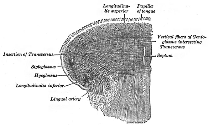
Fig. 1020. Coronal section Muscles of the Tongue
Showing intrinsic muscles supplied by the hypoglossal nerve. (Altered from Krause.)
The tongue is divided into lateral halves by a median fibrous septum which extends throughout its entire length and is fixed below to the hyoid bone. In either half there are two sets of muscles, extrinsic and intrinsic; the former have their origins outside the tongue, the latter are contained entirely within it.
- Longitudinalis linguæ superior (Superior lingualis) is a thin stratum of oblique and longitudinal fibers immediately underlying the mucous membrane on the dorsum of the tongue. It arises from the submucous fibrous layer close to the epiglottis and from the median fibrous septum, and runs forward to the edges of the tongue.
- Longitudinalis linguæ inferior (Inferior lingualis) is a narrow band situated on the under surface of the tongue between the Genioglossus and Hyoglossus. It extends from the root to the apex of the tongue: behind, some of its fibers are connected with the body of the hyoid bone; in front it blends with the fibers of the Styloglossus.
- Transversus linguæ (Transverse lingualis) consists of fibers which arise from the median fibrous septum and pass lateralward to be inserted into the submucous fibrous tissue at the sides of the tongue.
- Verticalis linguæ (Vertical lingualis) is found only at the borders of the forepart of the tongue. Its fibers extend from the upper to the under surface of the organ. The median fibrous septum of the tongue is very complete, so that the anastomosis between the two lingual arteries is not very free.
- Gray's Images: Development | Lymphatic | Neural | Vision | Hearing | Somatosensory | Integumentary | Respiratory | Gastrointestinal | Urogenital | Endocrine | Surface Anatomy | iBook | Historic Disclaimer
| Historic Disclaimer - information about historic embryology pages |
|---|
| Pages where the terms "Historic" (textbooks, papers, people, recommendations) appear on this site, and sections within pages where this disclaimer appears, indicate that the content and scientific understanding are specific to the time of publication. This means that while some scientific descriptions are still accurate, the terminology and interpretation of the developmental mechanisms reflect the understanding at the time of original publication and those of the preceding periods, these terms, interpretations and recommendations may not reflect our current scientific understanding. (More? Embryology History | Historic Embryology Papers) |
| iBook - Gray's Embryology | |
|---|---|

|
|
Reference
Gray H. Anatomy of the human body. (1918) Philadelphia: Lea & Febiger.
Cite this page: Hill, M.A. (2024, April 23) Embryology Gray1020.jpg. Retrieved from https://embryology.med.unsw.edu.au/embryology/index.php/File:Gray1020.jpg
- © Dr Mark Hill 2024, UNSW Embryology ISBN: 978 0 7334 2609 4 - UNSW CRICOS Provider Code No. 00098G
File history
Click on a date/time to view the file as it appeared at that time.
| Date/Time | Thumbnail | Dimensions | User | Comment | |
|---|---|---|---|---|---|
| current | 10:11, 11 May 2014 |  | 667 × 400 (53 KB) | Z8600021 (talk | contribs) | ==Fig. 1020. Coronal section of tongue== Showing intrinsic muscles. (Altered from Krause.) :'''Links:''' Tongue Development | Salivary Gland Development | Head Development | Musculoskeletal System Development | {{Gray Anatomy}} [[... |
| 10:11, 11 May 2014 |  | 667 × 400 (53 KB) | Z8600021 (talk | contribs) | ==Fig. 1020. Coronal section of tongue== Showing intrinsic muscles. (Altered from Krause.) :'''Links:''' Tongue Development | Salivary Gland Development | Head Development | Musculoskeletal System Development | {{Gray Anatomy}} [[... |
You cannot overwrite this file.
File usage
The following 2 pages use this file:

