File:Gray0919.jpg

Original file (500 × 731 pixels, file size: 93 KB, MIME type: image/jpeg)
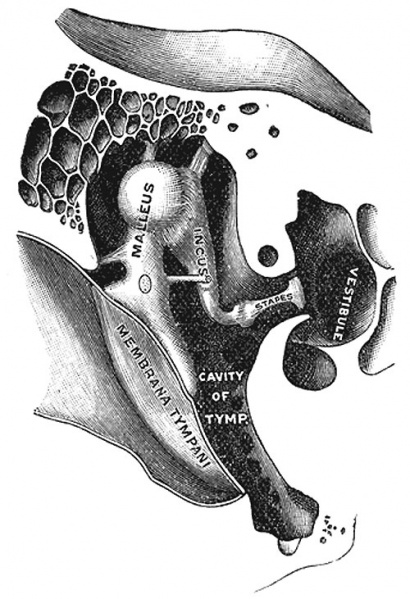
Chain of Ossicles and their Ligaments
Chain of ossicles and their ligaments, seen from the front in a vertical, transverse section of the tympanum. (Testut.)
Ligaments of the Ossicles (ligamenta ossiculorum auditus)
The ossicles are connected with the walls of the tympanic cavity by ligaments: three for the malleus, and one each for the incus and stapes.
- Anterior ligament of the malleus (lig. mallei anterius)
- attached by one end to the neck of the malleus, just above the anterior process, and by the other to the anterior wall of the tympanic cavity, close to the petrotympanic fissure, some of its fibers being prolonged through the fissure to reach the spina angularis of the sphenoid.
- Superior ligament of the malleus (lig. mallei superius)
- delicate, round bundle which descends from the roof of the epitympanic recess to the head of the malleus
- Lateral ligament of the malleus (lig. mallei laterale; external ligament of the malleus)
- triangular band passing from the posterior part of the notch of Rivinus to the head of the malleus
- Helmholtz described the anterior ligament and the posterior part of the lateral ligament as forming together the axis ligament around which the malleus rotates
- Posterior ligament of the incus (lig. incudis posterius)
- short, thick band connecting the end of the short crus of the incus to the fossa incudis
- Superior ligament of the incus (lig. incudis superius)
- has been described, but it is little more than a fold of mucous membrane
The vestibular surface and the circumference of the base of the stapes are covered with hyaline cartilage; that encircling the base is attached to the margin of the fenestra vestibuli by a fibrous ring, the annular ligament of the base of the stapes (lig. annulare baseos stapedis).
(Text modified from Gray's Anatomy)
- Gray's Images: Development | Lymphatic | Neural | Vision | Hearing | Somatosensory | Integumentary | Respiratory | Gastrointestinal | Urogenital | Endocrine | Surface Anatomy | iBook | Historic Disclaimer
| Historic Disclaimer - information about historic embryology pages |
|---|
| Pages where the terms "Historic" (textbooks, papers, people, recommendations) appear on this site, and sections within pages where this disclaimer appears, indicate that the content and scientific understanding are specific to the time of publication. This means that while some scientific descriptions are still accurate, the terminology and interpretation of the developmental mechanisms reflect the understanding at the time of original publication and those of the preceding periods, these terms, interpretations and recommendations may not reflect our current scientific understanding. (More? Embryology History | Historic Embryology Papers) |
| iBook - Gray's Embryology | |
|---|---|

|
|
Reference
Gray H. Anatomy of the human body. (1918) Philadelphia: Lea & Febiger.
Cite this page: Hill, M.A. (2024, April 20) Embryology Gray0919.jpg. Retrieved from https://embryology.med.unsw.edu.au/embryology/index.php/File:Gray0919.jpg
- © Dr Mark Hill 2024, UNSW Embryology ISBN: 978 0 7334 2609 4 - UNSW CRICOS Provider Code No. 00098G
File history
Click on a date/time to view the file as it appeared at that time.
| Date/Time | Thumbnail | Dimensions | User | Comment | |
|---|---|---|---|---|---|
| current | 03:16, 20 May 2011 |  | 500 × 731 (93 KB) | S8600021 (talk | contribs) | Chain of ossicles and their ligaments, seen from the front in a vertical, transverse section of the tympanum. (Testut.) ===Ligaments of the Ossicles (ligamenta ossiculorum auditus)=== The ossicles are connected with the walls of the tympanic cavity by |
You cannot overwrite this file.
File usage
The following 2 pages use this file:
