File:Gray0894.jpg
Gray0894.jpg (710 × 400 pixels, file size: 74 KB, MIME type: image/jpeg)
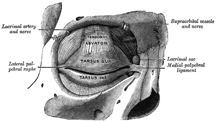
The tarsi and their ligaments
Right eye; front view.
The tarsi (tarsal plates) (Fig. 894) are two thin, elongated plates of dense connective tissue, about 2.5 cm. in length; one is placed in each eyelid, and contributes to its form and support. The superior tarsus (tarsus superior; superior tarsal plate), the larger, is of a semilunar form, about 10 mm. in breadth at the center, and gradually narrowing toward its extremities. To the anterior surface of this plate the aponeurosis of the Levator palpebræ superioris is attached. The inferior tarsus (tarsus inferior; inferior tarsal plate), the smaller, is thin, elliptical in form, and has a vertical diameter of about 5 mm. The free or ciliary margins of these plates are thick and straight. The attached or orbital margins are connected to the circumference of the orbit by the orbital septum. The lateral angles are attached to the zygomatic bone by the lateral palpebral raphé. The medial angles of the two plates end at the lacus lacrimalis, and are attached to the frontal process of the maxilla by the medial palpebral ligament (page 381).
(Text modified from Gray's 1918 Anatomy)
- Gray's Images: Development | Lymphatic | Neural | Vision | Hearing | Somatosensory | Integumentary | Respiratory | Gastrointestinal | Urogenital | Endocrine | Surface Anatomy | iBook | Historic Disclaimer
| Historic Disclaimer - information about historic embryology pages |
|---|
 |
| iBook - Gray's Embryology | |
|---|---|

|
|
Reference
Gray H. Anatomy of the human body. (1918) Philadelphia: Lea & Febiger.
Cite this page: Hill, M.A. (2024, April 19) Embryology Gray0894.jpg. Retrieved from https://embryology.med.unsw.edu.au/embryology/index.php/File:Gray0894.jpg
- © Dr Mark Hill 2024, UNSW Embryology ISBN: 978 0 7334 2609 4 - UNSW CRICOS Provider Code No. 00098G
File history
Click on a date/time to view the file as it appeared at that time.
| Date/Time | Thumbnail | Dimensions | User | Comment | |
|---|---|---|---|---|---|
| current | 22:57, 19 August 2012 | | 710 × 400 (74 KB) | Z8600021 (talk | contribs) | ==The tarsi and their ligaments== Right eye; front view. The tarsi (tarsal plates) (Fig. 894) are two thin, elongated plates of dense connective tissue, about 2.5 cm. in length; one is placed in each eyelid, and contributes to its form and support. The |
You cannot overwrite this file.
File usage
The following 2 pages use this file: