File:Gray0889.jpg
Gray0889.jpg (800 × 519 pixels, file size: 103 KB, MIME type: image/jpeg)
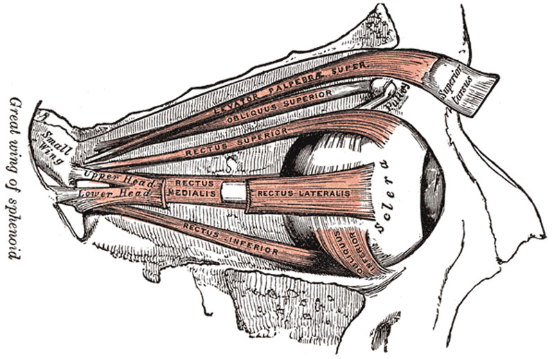
Muscles of the right orbit
The Levator palpebræ superioris (Fig. 888) is thin, flat, and triangular in shape. It arises from the under surface of the small wing of the sphenoid, above and in front of the optic foramen, from which it is separated by the origin of the Rectus superior. At its origin, it is narrow and tendinous, but soon becomes broad and fleshy, and ends anteriorly in a wide aponeurosis which splits into three lamellæ. The superficial lamella blends with the upper part of the orbital septum, and is prolonged forward above the superior tarsus to the palpebral part of the Orbicularis oculi, and to the deep surface of the skin of the upper eyelid. The middle lamella, largely made up of non-striped muscular fibers, is inserted into the upper margin of the superior tarsus, while the deepest lamella blends with an expansion from the sheath of the Rectus superior and with it is attached to the superior fornix of the conjunctiva.
Whitnall has pointed out that the upper part of the sheath of the Levator palpebræ becomes thickened in front and forms, above the anterior part of the muscle, a transverse ligamentous band which is attached to the sides of the orbital cavity. On the medial side it is mainly fixed to the pulley of the Obliquus superior, but some fibers are attached to the bone behind the pulley and a slip passes forward and bridges over the supraorbital notch; on the lateral side it is fixed to the capsule of the lacrimal gland and to the frontal bone. In front of the transverse ligamentous band the sheath is continued over the aponeurosis of the Levator palpebræ, as a thin connective-tissue layer which is fixed to the upper orbital margin immediatly behind the attachment of the orbital septum. When the Levator palpebræ contracts, the lateral and medial parts of the ligamentous band are stretched and check the action of the muscle; the retraction of the upper eyelid is checked also by the orbital septum coming into contact with the transverse part of the ligamentous band.
The four Recti (Fig. 889) arise from a fibrous ring (annulus tendineus communis) which surrounds the upper, medial, and lower margins of the optic foramen and encircles the optic nerve (Fig. 890). The ring is completed by a tendinous bridge prolonged over the lower and medial part of the superior orbital fissure and attached to a tubercle on the margin of the great wing of the sphenoid, bounding the fissure. Two specialized parts of this fibrous ring may be made out: a lower, the ligament or tendon of Zinn, which gives origin to the Rectus inferior, part of the Rectus internus, and the lower head of origin of the Rectus lateralis; and an upper, which gives origin to the Rectus superior, the rest of the Rectus medialis, and the upper head of the Rectus lateralis. This upper band is sometimes termed the superior tendon of Lockwood. Each muscle passes forward in the position implied by its name, to be inserted by a tendinous expansion into the sclera, about 6 mm. from the margin of the cornea. Between the two heads of the Rectus lateralis is a narrow interval, through which pass the two divisions of the oculomotor nerve, the nasociliary nerve, the abducent nerve, and the ophthalmic vein. Although these muscles present a common origin and are inserted in a similar manner into the sclera, there are certain differences to be observed in them as regards their length and breadth. The Rectus medialis is the broadest, the Rectus lateralis the longest, and the Rectus superior the thinnest and narrowest.
The Obliquus oculi superior (superior oblique) is a fusiform muscle, placed at the upper and medial side of the orbit. It arises immediately above the margin of the optic foramen, above and medial to the origin of the Rectus superior, and, passing forward, ends in a rounded tendon, which plays in a fibrocartilaginous ring or pulley attached to the trochlear fovea of the frontal bone. The contiguous surfaces of the tendon and ring are lined by a delicate mucous sheath, and enclosed in a thin fibrous investment. The tendon is reflected backward, lateralward, and downward beneath the Rectus superior to the lateral part of the bulb of the eye, and is inserted into the sclera, behind the equator of the eyeball, the insertion of the muscle lying between the Rectus superior and Rectus lateralis.
The Obliquus oculi inferior (inferior oblique) is a thin, narrow muscle, placed near the anterior margin of the floor of the orbit. It arises from the orbital surface of the maxilla, lateral to the lacrimal groove. Passing lateralward, backward, and upward, at first between the Rectus inferior and the floor of the orbit, and then between the bulb of the eye and the Rectus lateralis, it is inserted into the lateral part of the sclera between the Rectus superior and Rectus lateralis, near to, but somewhat behind the insertion of the Obliquus superior.
(Text modified from Gray's 1918 Anatomy)
- Gray's Images: Development | Lymphatic | Neural | Vision | Hearing | Somatosensory | Integumentary | Respiratory | Gastrointestinal | Urogenital | Endocrine | Surface Anatomy | iBook | Historic Disclaimer
| Historic Disclaimer - information about historic embryology pages |
|---|
 |
| iBook - Gray's Embryology | |
|---|---|

|
|
Reference
Gray H. Anatomy of the human body. (1918) Philadelphia: Lea & Febiger.
Cite this page: Hill, M.A. (2024, April 23) Embryology Gray0889.jpg. Retrieved from https://embryology.med.unsw.edu.au/embryology/index.php/File:Gray0889.jpg
- © Dr Mark Hill 2024, UNSW Embryology ISBN: 978 0 7334 2609 4 - UNSW CRICOS Provider Code No. 00098G
File history
Click on a date/time to view the file as it appeared at that time.
| Date/Time | Thumbnail | Dimensions | User | Comment | |
|---|---|---|---|---|---|
| current | 22:41, 19 August 2012 | | 800 × 519 (103 KB) | Z8600021 (talk | contribs) | ==Muscles of the right orbit== The Levator palpebræ superioris (Fig. 888) is thin, flat, and triangular in shape. It arises from the under surface of the small wing of the sphenoid, above and in front of the optic foramen, from which it is separated by |
You cannot overwrite this file.
File usage
The following page uses this file: