File:Gray0886.jpg
Gray0886.jpg (500 × 368 pixels, file size: 17 KB, MIME type: image/jpeg)
Lens Profile
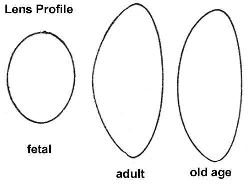
Profile views of the lens at different periods of life. 1. In the fetus. 2. In adult life. 3. In old age.
In the fetus, the lens is nearly spherical, and has a slightly reddish tint; it is soft and breaks down readily on the slightest pressure. A small branch from the arteria centralis retinæ runs forward, as already mentioned, through the vitreous body to the posterior part of the capsule of the lens, where its branches radiate and form a plexiform network, which covers the posterior surface of the capsule, and they are continuous around the margin of the capsule with the vessels of the pupillary membrane, and with those of the iris. In the adult, the lens is colorless, transparent, firm in texture, and devoid of vessels. In old age it becomes flattened on both surfaces, slightly opaque, of an amber tint, and increased in density (Fig. 886).
(Text modified from Gray's 1918 Anatomy)
- Gray's Images: Development | Lymphatic | Neural | Vision | Hearing | Somatosensory | Integumentary | Respiratory | Gastrointestinal | Urogenital | Endocrine | Surface Anatomy | iBook | Historic Disclaimer
| Historic Disclaimer - information about historic embryology pages |
|---|
| Pages where the terms "Historic" (textbooks, papers, people, recommendations) appear on this site, and sections within pages where this disclaimer appears, indicate that the content and scientific understanding are specific to the time of publication. This means that while some scientific descriptions are still accurate, the terminology and interpretation of the developmental mechanisms reflect the understanding at the time of original publication and those of the preceding periods, these terms, interpretations and recommendations may not reflect our current scientific understanding. (More? Embryology History | Historic Embryology Papers) |
| iBook - Gray's Embryology | |
|---|---|

|
|
Reference
Gray H. Anatomy of the human body. (1918) Philadelphia: Lea & Febiger.
Cite this page: Hill, M.A. (2024, April 23) Embryology Gray0886.jpg. Retrieved from https://embryology.med.unsw.edu.au/embryology/index.php/File:Gray0886.jpg
- © Dr Mark Hill 2024, UNSW Embryology ISBN: 978 0 7334 2609 4 - UNSW CRICOS Provider Code No. 00098G
File history
Click on a date/time to view the file as it appeared at that time.
| Date/Time | Thumbnail | Dimensions | User | Comment | |
|---|---|---|---|---|---|
| current | 22:30, 19 August 2012 |  | 500 × 368 (17 KB) | Z8600021 (talk | contribs) | ==Lens Profile== Profile views of the lens at different periods of life. 1. In the fetus. 2. In adult life. 3. In old age. (Text modified from Gray's 1918 Anatomy) {{Gray Anatomy}} Category:Cartoon Category:Senses Category:Vision [[Categor |
You cannot overwrite this file.
File usage
The following page uses this file:

