File:Gray0865.jpg
Gray0865.jpg (759 × 400 pixels, file size: 70 KB, MIME type: image/jpeg)
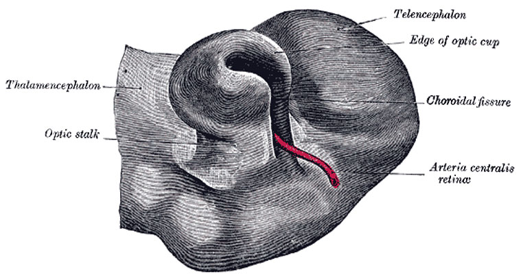
Human Optic Cup and Choroidal Fissure
Optic cup and choroidal fissure seen from below, from a human embryo of about four weeks. (Kollmann.)
Development
The eyes begin to develop as a pair of diverticula from the lateral aspects of the forebrain. These diverticula make their appearance before the closure of the anterior end of the neural tube; after the closure of the tube they are known as the optic vesicles. They project toward the sides of the head, and the peripheral part of each expands to form a hollow bulb, while the proximal part remains narrow and constitutes the optic stalk (Figs. 863, 864). The ectoderm overlying the bulb becomes thickened, invaginated, and finally severed from the ectodermal covering of the head as a vesicle of cells, the lens vesicle, which constitutes the rudiment of the crystalline lens. The outer wall of the bulb becomes thickened and invaginated, and the bulb is thus converted into a cup, the optic cup, consisting of two strata of cells (Fig. 864). These two strata are continuous with each other at the cup margin, which ultimately overlaps the front of the lens and reaches as far forward as the future aperture of the pupil. The invagination is not limited to the outer wall of the bulb, but involves also its postero-inferior surface and extends in the form of a groove for some distance along the optic stalk, so that, for a time, a gap or fissure, the choroidal fissure, exists in the lower part of the cup (Fig. 865). Through the groove and fissure the mesoderm extends into the optic stalk and cup, and in this mesoderm a bloodvessel is developed; during the seventh week the groove and fissure are closed and the vessel forms the central artery of the retina. Sometimes the choroidal fissure persists, and when this occurs the choroid and iris in the region of the fissure remain undeveloped, giving rise to the condition known as coloboma of the choroid or iris.
(Text modified from Gray's 1918 Anatomy)
- Gray's Images: Development | Lymphatic | Neural | Vision | Hearing | Somatosensory | Integumentary | Respiratory | Gastrointestinal | Urogenital | Endocrine | Surface Anatomy | iBook | Historic Disclaimer
| Historic Disclaimer - information about historic embryology pages |
|---|
| Pages where the terms "Historic" (textbooks, papers, people, recommendations) appear on this site, and sections within pages where this disclaimer appears, indicate that the content and scientific understanding are specific to the time of publication. This means that while some scientific descriptions are still accurate, the terminology and interpretation of the developmental mechanisms reflect the understanding at the time of original publication and those of the preceding periods, these terms, interpretations and recommendations may not reflect our current scientific understanding. (More? Embryology History | Historic Embryology Papers) |
| iBook - Gray's Embryology | |
|---|---|

|
|
Reference
Gray H. Anatomy of the human body. (1918) Philadelphia: Lea & Febiger.
Cite this page: Hill, M.A. (2024, April 20) Embryology Gray0865.jpg. Retrieved from https://embryology.med.unsw.edu.au/embryology/index.php/File:Gray0865.jpg
- © Dr Mark Hill 2024, UNSW Embryology ISBN: 978 0 7334 2609 4 - UNSW CRICOS Provider Code No. 00098G
File history
Click on a date/time to view the file as it appeared at that time.
| Date/Time | Thumbnail | Dimensions | User | Comment | |
|---|---|---|---|---|---|
| current | 08:51, 19 August 2012 |  | 759 × 400 (70 KB) | Z8600021 (talk | contribs) | ==Human Optic Cup and Choroidal Fissure== Optic cup and choroidal fissure seen from below, from a human embryo of about four weeks. (Kollmann.) (Text modified from Gray's 1918 Anatomy) {{Gray Anatomy}} Category:Cartoon Category:Senses [[Categ |
You cannot overwrite this file.
File usage
The following page uses this file:

