File:Gray0859.jpg
Gray0859.jpg (694 × 600 pixels, file size: 114 KB, MIME type: image/jpeg)
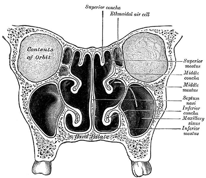
Fig. 859. Coronal section of Nasal Cavities
Ethmoidal Air Cells
(cellulæ ethmoidales) consist of numerous thin-walled cavities situated in the ethmoidal labyrinth and completed by the frontal, maxilla, lacrimal, sphenoidal, and palatine. They lie between the upper parts of the nasal cavities and the orbits, and are separated from these cavities by thin bony laminæ. On either side they are arranged in three groups, anterior, middle, and posterior. The anterior and middle groups open into the middle meatus of the nose, the former by way of the infundibulum, the latter on or above the bulla ethmoidalis. The posterior cells open into the superior meatus under cover of the superior nasal concha; sometimes one or more opens into the sphenoidal sinus. The ethmoidal cells begin to develop during fetal life.
Maxillary Sinus
(sinus maxillaris; antrum of Highmore), the largest of the accessory sinuses of the nose, is a pyramidal cavity in the body of the maxilla. Its base is formed by the lateral wall of the nasal cavity, and its apex extends into the zygomatic process. Its roof or orbital wall is frequently ridged by the infra-orbital canal, while its floor is formed by the alveolar process and is usually 1/2 to 10 mm. below the level of the floor of the nose; projecting into the floor are several conical elevations corresponding with the roots of the first and second molar teeth, and in some cases the floor is perforated by one or more of these roots. The size of the sinus varies in different skulls, and even on the two sides of the same skull. The adult capacity varies from 9.5 c.c. to 20 c.c., average about 14.75 c.c. The following measurements are those of an average-sized sinus: vertical height opposite the first molar tooth, 3.75 cm.; transverse breadth, 2.5 cm.; antero-posterior depth, 3 cm. In the antero-superior part of its base is an opening through which it communicates with the lower part of the hiatus semilunaris; a second orifice is frequently seen in, or immediately behind, the hiatus. The maxillary sinus appears as a shallow groove on the medial surface of the bone about the fourth month of fetal life, but does not reach its full size until after the second dentition. 142 At birth it measures about 7 mm. in the dorso-ventral direction and at twenty months about 20 mm.
- Gray's Images: Development | Lymphatic | Neural | Vision | Hearing | Somatosensory | Integumentary | Respiratory | Gastrointestinal | Urogenital | Endocrine | Surface Anatomy | iBook | Historic Disclaimer
| Historic Disclaimer - information about historic embryology pages |
|---|
| Pages where the terms "Historic" (textbooks, papers, people, recommendations) appear on this site, and sections within pages where this disclaimer appears, indicate that the content and scientific understanding are specific to the time of publication. This means that while some scientific descriptions are still accurate, the terminology and interpretation of the developmental mechanisms reflect the understanding at the time of original publication and those of the preceding periods, these terms, interpretations and recommendations may not reflect our current scientific understanding. (More? Embryology History | Historic Embryology Papers) |
| iBook - Gray's Embryology | |
|---|---|

|
|
Reference
Gray H. Anatomy of the human body. (1918) Philadelphia: Lea & Febiger.
Cite this page: Hill, M.A. (2024, April 24) Embryology Gray0859.jpg. Retrieved from https://embryology.med.unsw.edu.au/embryology/index.php/File:Gray0859.jpg
- © Dr Mark Hill 2024, UNSW Embryology ISBN: 978 0 7334 2609 4 - UNSW CRICOS Provider Code No. 00098G
File history
Click on a date/time to view the file as it appeared at that time.
| Date/Time | Thumbnail | Dimensions | User | Comment | |
|---|---|---|---|---|---|
| current | 14:32, 3 May 2013 |  | 694 × 600 (114 KB) | Z8600021 (talk | contribs) | ==Fig. 859. Coronal section of nasal cavities== {{Gray Anatomy}} Category:Historic Embryology Category:Gray's 1918 Anatomy Category:Cartoon Category:Senses Category:Smell Category:Respiratory |
You cannot overwrite this file.
File usage
The following 2 pages use this file:

