File:Gray0806.jpg
Original file (600 × 771 pixels, file size: 173 KB, MIME type: image/jpeg)
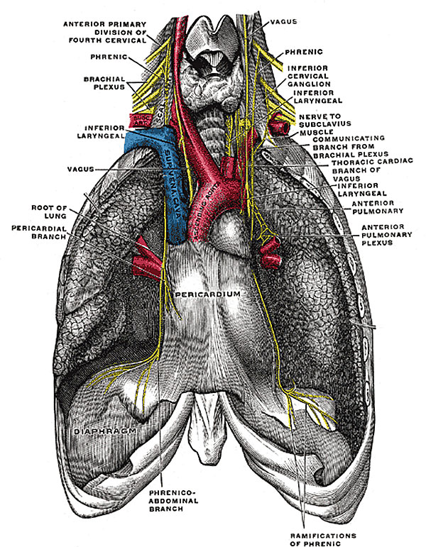
Fig. 806. The phrenic nerve and its relations with the vagus nerve
The Phrenic Nerve (n. phrenicus; internal respiratory nerve of Bell) contains motor and sensory fibers in the proportion of about two to one. It arises chiefly from the fourth cervical nerve, but receives a branch from the third and another from the fifth; (the fibers from the fifth occasionally come through the nerve to the Subclavius.) It descends to the root of the neck, running obliquely across the front of the Scalenus anterior, and beneath the Sternocleidomastoideus, the inferior belly of the Omohyoideus, and the transverse cervical and transverse scapular vessels. It next passes in front of the first part of the subclavian artery, between it and the subclavian vein, and, as it enters the thorax, crosses the internal mammary artery near its origin. Within the thorax, it descends nearly vertically in front of the root of the lung, and then between the pericardium and the mediastinal pleura, to the diaphragm, where it divides into branches, which pierce that muscle, and are distributed to its under surface. In the thorax it is accompanied by the pericardiacophrenic branch of the internal mammary artery.
The two phrenic nerves differ in their length, and also in their relations at the upper part of the thorax.
- The right nerve is situated more deeply, and is shorter and more vertical in direction than the left; it lies lateral to the right innominate vein and superior vena cava.
- The left nerve is rather longer than the right, from the inclination of the heart to the left side, and from the diaphragm being lower on this than on the right side. At the root of the neck it is crossed by the thoracic duct; in the superior mediastinal cavity it lies between the left common carotid and left subclavian arteries, and crosses superficial to the vagus on the left side of the arch of the aorta.
- Gray's Images: Development | Lymphatic | Neural | Vision | Hearing | Somatosensory | Integumentary | Respiratory | Gastrointestinal | Urogenital | Endocrine | Surface Anatomy | iBook | Historic Disclaimer
| Historic Disclaimer - information about historic embryology pages |
|---|
 |
| iBook - Gray's Embryology | |
|---|---|

|
|
Reference
Gray H. Anatomy of the human body. (1918) Philadelphia: Lea & Febiger.
Cite this page: Hill, M.A. (2024, April 24) Embryology Gray0806.jpg. Retrieved from https://embryology.med.unsw.edu.au/embryology/index.php/File:Gray0806.jpg
- © Dr Mark Hill 2024, UNSW Embryology ISBN: 978 0 7334 2609 4 - UNSW CRICOS Provider Code No. 00098G
File history
Click on a date/time to view the file as it appeared at that time.
| Date/Time | Thumbnail | Dimensions | User | Comment | |
|---|---|---|---|---|---|
| current | 15:26, 24 August 2010 | | 600 × 771 (173 KB) | S8600021 (talk | contribs) | The phrenic nerve and its relations with the vagus nerve. |
You cannot overwrite this file.
File usage
The following page uses this file: