File:Gray0788.jpg
Original file (815 × 750 pixels, file size: 91 KB, MIME type: image/jpeg)
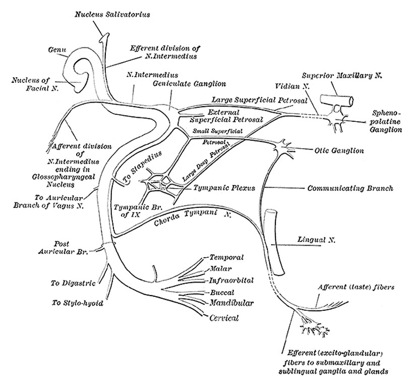
Fig. 788. Plan of the Facial and Intermediate Nerves and their Communication with Other Nerves
(N. Facialis; Seventh Nerve; CN VII)
The facial nerve (Figs. 788, 790) consists of a motor and a sensory part, the latter being frequently described under the name of the nervus intermedius (pars intermedii of Wrisberg) (Fig. 788). The two parts emerge at the lower border of the pons in the recess between the olive and the inferior peduncle, the motor part being the more medial, immediately to the lateral side of the sensory part is the acoustic nerve.
The motor part supplies somatic motor fibers to the muscles of the face, scalp, and auricle, the Buccinator and Platysma, the Stapedius, the Stylohyoideus, and posterior belly of the Digastricus; it also contains some sympathetic motor fibers which constitute the vasodilator nerves of the submaxillary and sublingual glands, and are conveyed through the chorda tympani nerve. These are preganglionic fibers of the sympathetic system and terminate in the submaxillary ganglion and small ganglia in the hilus of the submaxillary gland. From these ganglia postganglionic fibers are conveyed to these glands. The sensory part contains the fibers of taste for the anterior two-thirds of the tongue and a few somatic sensory fibers from the middle ear region. A few splanchnic sensory fibers are also present.
Motor Root
Arises from a nucleus which lies deeply in the reticular formation of the lower part of the pons. This nucleus is situated above the nucleus ambiguus, behind the superior olivary nucleus, and medial to the spinal tract of the trigeminal nerve. From this origin the fibers pursue a curved course in the substance of the pons. They first pass backward and medialward toward the rhomboid fossa, and, reaching the posterior end of the nucleus of the abducent nerve, run upward close to the middle line beneath the colliculus fasciculus. At the anterior end of the nucleus of the abducent nerve they make a second bend, and run downward and forward through the pons to their point of emergence between the olive and the inferior peduncle.
Sensory root
Arises from the genicular ganglion, which is situated on the geniculum of the facial nerve in the facial canal, behind the hiatus of the canal. The cells of this ganglion are unipolar, and the single process divides in a T-shaped manner into central and peripheral branches. The central branches leave the trunk of the facial nerve in the internal acoustic meatus, and form the sensory root; the peripheral branches are continued into the chorda tympani and greater superficial petrosal nerves. Entering the brain at the lower border of the pons between the motor root and the acoustic nerve, the fibers of the sensory root pass into the substance of the medulla oblongata and end in the upper part of the terminal nucleus of the glossopharyngeal nerve and in the fasciculus solitarius.
(text modified from Gray's Anatomy)
- Gray's Images: Development | Lymphatic | Neural | Vision | Hearing | Somatosensory | Integumentary | Respiratory | Gastrointestinal | Urogenital | Endocrine | Surface Anatomy | iBook | Historic Disclaimer
| Historic Disclaimer - information about historic embryology pages |
|---|
 |
| iBook - Gray's Embryology | |
|---|---|

|
|
Reference
Gray H. Anatomy of the human body. (1918) Philadelphia: Lea & Febiger.
Cite this page: Hill, M.A. (2024, April 18) Embryology Gray0788.jpg. Retrieved from https://embryology.med.unsw.edu.au/embryology/index.php/File:Gray0788.jpg
- © Dr Mark Hill 2024, UNSW Embryology ISBN: 978 0 7334 2609 4 - UNSW CRICOS Provider Code No. 00098G
File history
Click on a date/time to view the file as it appeared at that time.
| Date/Time | Thumbnail | Dimensions | User | Comment | |
|---|---|---|---|---|---|
| current | 13:00, 15 May 2013 | | 815 × 750 (91 KB) | Z8600021 (talk | contribs) | ==Fig. 788. Plan of the Facial and Intermediate Nerves and their Communication with Other Nerves== (N. Facialis; Seventh Nerve; CN VII) The facial nerve (Figs. 788, 790) consists of a motor and a sensory part, the l... |
You cannot overwrite this file.
File usage
The following 3 pages use this file: