File:Gray0784.jpg
Original file (851 × 600 pixels, file size: 137 KB, MIME type: image/jpeg)
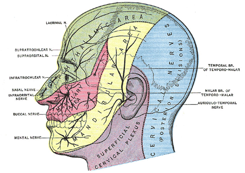
Fig. 784. Sensory Areas of the Head
Showing the general distribution of the three divisions of the fifth nerve (N. Trigeminus, Trifacial Nerve, CN V).
The trigeminal nerve is the largest cranial nerve and is the great sensory nerve of the head and face, and the motor nerve of the muscles of mastication. It emerges from the side of the pons, near its upper border, by a small motor and a large sensory root—the former being situated in front of and medial to the latter.
Ophthalmic Nerve
(n. ophthalmicus) (Figs. 776, 777), or first division of the trigeminal, is a sensory nerve. It supplies branches to the cornea, ciliary body, and iris; to the lacrimal gland and conjunctiva; to the part of the mucous membrane of the nasal cavity; and to the skin of the eyelids, eyebrow, forehead, and nose. It is the smallest of the three divisions of the trigeminal, and arises from the upper part of the semilunar ganglion as a short, flattened band, about 2.5 cm. long, which passes forward along the lateral wall of the cavernous sinus, below the oculomotor and trochlear nerves; just before entering the orbit, through the superior orbital fissure, it divides into three branches, lacrimal, frontal, and nasociliary.
Maxillary Nerve
(n. maxillaris; superior maxillary nerve) ( Fig. 778), or second division of the trigeminal, is a sensory nerve. It is intermediate, both in position and size, between the ophthalmic and mandibular. It begins at the middle of the semilunar ganglion as a flattened plexiform band, and, passing horizontally forward, it leaves the skull through the foramen rotundum, where it becomes more cylindrical in form, and firmer in texture. It then crosses the pterygopalatine fossa, inclines lateralward on the back of the maxilla, and enters the orbit through the inferior orbital fissure; it traverses the infraorbital groove and canal in the floor of the orbit, and appears upon the face at the infraorbital foramen. At its termination, the nerve lies beneath the Quadratus labii superioris, and divides into a leash of branches which spread out upon the side of the nose, the lower eyelid, and the upper lip, joining with filaments of the facial nerve.
Mandibular Nerve
(n. mandibularis; inferior maxillary nerve) (Figs. 778, 781) supplies the teeth and gums of the mandible, the skin of the temporal region, the auricula, the lower lip, the lower part of the face, and the muscles of mastication; it also supplies the mucous membrane of the anterior two-thirds of the tongue. It is the largest of the three divisions of the fifth, and is made up of two roots: a large, sensory root proceeding from the inferior angle of the semilunar ganglion, and a small motor root (the motor part of the trigeminal), which passes beneath the ganglion, and unites with the sensory root, just after its exit through the foramen ovale. Immediately beneath the base of the skull, the nerve gives off from its medial side a recurrent branch (nervus spinosus) and the nerve to the Pterygoideus internus, and then divides into two trunks, an anterior and a posterior.
(Image Modified from Testut.)
(text modified from Gray's Anatomy)
- Gray's Images: Development | Lymphatic | Neural | Vision | Hearing | Somatosensory | Integumentary | Respiratory | Gastrointestinal | Urogenital | Endocrine | Surface Anatomy | iBook | Historic Disclaimer
| Historic Disclaimer - information about historic embryology pages |
|---|
 |
| iBook - Gray's Embryology | |
|---|---|

|
|
Reference
Gray H. Anatomy of the human body. (1918) Philadelphia: Lea & Febiger.
Cite this page: Hill, M.A. (2024, April 25) Embryology Gray0784.jpg. Retrieved from https://embryology.med.unsw.edu.au/embryology/index.php/File:Gray0784.jpg
- © Dr Mark Hill 2024, UNSW Embryology ISBN: 978 0 7334 2609 4 - UNSW CRICOS Provider Code No. 00098G
File history
Click on a date/time to view the file as it appeared at that time.
| Date/Time | Thumbnail | Dimensions | User | Comment | |
|---|---|---|---|---|---|
| current | 13:13, 15 May 2013 | | 851 × 600 (137 KB) | Z8600021 (talk | contribs) | ==Fig. 784. Sensory Areas of the Head== Showing the general distribution of the three divisions of the fifth nerve (N. Trigeminus, Trifacial Nerve, CN V). The trigeminal nerve is the largest cranial nerve and is the great sensory nerve of the head a... |
You cannot overwrite this file.
File usage
The following 3 pages use this file: