File:Gray0770.jpg
Gray0770.jpg (700 × 275 pixels, file size: 68 KB, MIME type: image/jpeg)
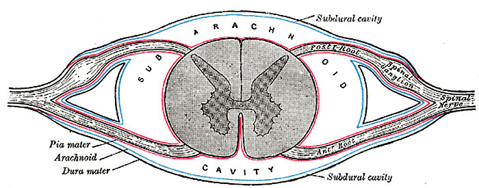
Fig. 770. Diagrammatic transverse section of the medulla spinalis and its membranes
The Spinal Pia Mater (pia mater spinalis; pia of the cord) (Fig. 767, Fig. 770) is thicker, firmer, and less vascular than the cranial pia mater: this is due to the fact that it consists of two layers, the outer or additional one being composed of bundles of connective-tissue fibers, arranged for the most part longitudinally. Between the layers are cleft-like spaces which communicate with the subarachnoid cavity, and a number of bloodvessels which are enclosed in perivascular lymphatic sheaths. The spinal pia mater covers the entire surface of the medulla spinalis, and is very intimately adherent to it; in front it sends a process backward into the anterior fissure. A longitudinal fibrous band, called the linea splendens, extends along the middle line of the anterior surface; and a somewhat similar band, the ligamentum denticulatum, is situated on either side. Below the conus medullaris, the pia mater is continued as a long, slender filament (filum terminale), which descends through the center of the mass of nerves forming the cauda equina. It blends with the dura mater at the level of the lower border of the second sacral vertebra, and extends downward as far as the base of the coccyx, where it fuses with the periosteum. It assists in maintaining the medulla spinalis in its position during the movements of the trunk, and is, from this circumstance, called the central ligament of the medulla spinals.
(text modified from Gray's Anatomy)
- Gray's Images: Development | Lymphatic | Neural | Vision | Hearing | Somatosensory | Integumentary | Respiratory | Gastrointestinal | Urogenital | Endocrine | Surface Anatomy | iBook | Historic Disclaimer
| Historic Disclaimer - information about historic embryology pages |
|---|
| Pages where the terms "Historic" (textbooks, papers, people, recommendations) appear on this site, and sections within pages where this disclaimer appears, indicate that the content and scientific understanding are specific to the time of publication. This means that while some scientific descriptions are still accurate, the terminology and interpretation of the developmental mechanisms reflect the understanding at the time of original publication and those of the preceding periods, these terms, interpretations and recommendations may not reflect our current scientific understanding. (More? Embryology History | Historic Embryology Papers) |
| iBook - Gray's Embryology | |
|---|---|

|
|
Reference
Gray H. Anatomy of the human body. (1918) Philadelphia: Lea & Febiger.
Cite this page: Hill, M.A. (2024, April 25) Embryology Gray0770.jpg. Retrieved from https://embryology.med.unsw.edu.au/embryology/index.php/File:Gray0770.jpg
- © Dr Mark Hill 2024, UNSW Embryology ISBN: 978 0 7334 2609 4 - UNSW CRICOS Provider Code No. 00098G
File history
Click on a date/time to view the file as it appeared at that time.
| Date/Time | Thumbnail | Dimensions | User | Comment | |
|---|---|---|---|---|---|
| current | 10:26, 6 February 2016 | 700 × 275 (68 KB) | Z8600021 (talk | contribs) |
You cannot overwrite this file.
File usage
The following page uses this file:

