File:Gray0704.jpg
From Embryology
Gray0704.jpg (800 × 555 pixels, file size: 83 KB, MIME type: image/jpeg)
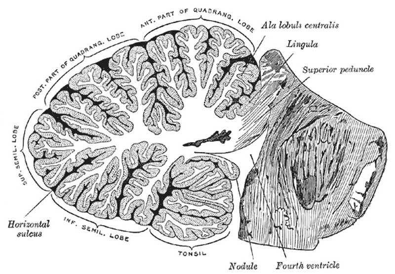
Fig. 704. Cerebellum
Sagittal section of the cerebellum, near the junction of the vermis with the hemisphere. (Schäfer.)
- constitutes the largest part of the hindbrain.
- lies behind the pons and medulla oblongata
- between its central portion and these structures is the cavity of the fourth ventricle.
- rests on the inferior occipital fossæ, while above it is the tentorium cerebella
- a fold of dura mater separates it from the tentorial surface of the cerebrum.
- somewhat oval in form, but constricted medially and flattened from above downward, its greatest diameter being from side.
- surface is not convoluted like that of the cerebrum
- traversed by numerous curved furrows or sulci, which vary in depth at different parts, and separate the laminæ of which it is composed.
- average weight in the male is about 150 gms.
Proportion between the cerebellum and cerebrum
- infant about 1 to 20.
- adult about 1 to 8.
- Cerebellum Images: Anatomical position | Upper surface | Projection fibres | Sagittal section | Transverse section folium | Cerebellum Development
- Gray's Images: Development | Lymphatic | Neural | Vision | Hearing | Somatosensory | Integumentary | Respiratory | Gastrointestinal | Urogenital | Endocrine | Surface Anatomy | iBook | Historic Disclaimer
| Historic Disclaimer - information about historic embryology pages |
|---|
| Pages where the terms "Historic" (textbooks, papers, people, recommendations) appear on this site, and sections within pages where this disclaimer appears, indicate that the content and scientific understanding are specific to the time of publication. This means that while some scientific descriptions are still accurate, the terminology and interpretation of the developmental mechanisms reflect the understanding at the time of original publication and those of the preceding periods, these terms, interpretations and recommendations may not reflect our current scientific understanding. (More? Embryology History | Historic Embryology Papers) |
| iBook - Gray's Embryology | |
|---|---|

|
|
Reference
Gray H. Anatomy of the human body. (1918) Philadelphia: Lea & Febiger.
Cite this page: Hill, M.A. (2024, April 24) Embryology Gray0704.jpg. Retrieved from https://embryology.med.unsw.edu.au/embryology/index.php/File:Gray0704.jpg
- © Dr Mark Hill 2024, UNSW Embryology ISBN: 978 0 7334 2609 4 - UNSW CRICOS Provider Code No. 00098G
File history
Click on a date/time to view the file as it appeared at that time.
| Date/Time | Thumbnail | Dimensions | User | Comment | |
|---|---|---|---|---|---|
| current | 13:43, 7 December 2010 |  | 800 × 555 (83 KB) | S8600021 (talk | contribs) | ==Cerebellum== Sagittal section of the cerebellum, near the junction of the vermis with the hemisphere. (Schäfer.) {{Template:Gray Anatomy}} Category:Neural |
You cannot overwrite this file.

