File:Gray0678.jpg
Gray0678.jpg (600 × 598 pixels, file size: 35 KB, MIME type: image/jpeg)
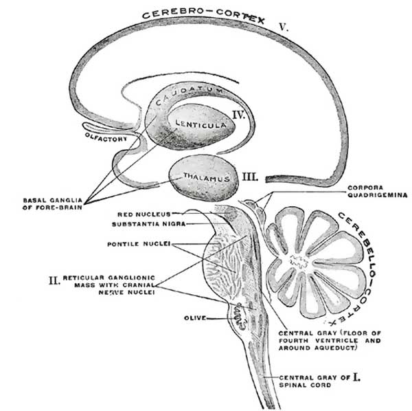
Fig. 678. Brain - Schematic representation of the chief ganglionic categories (I to V).
(Spitzka.)
General Considerations and Divisions
The brain, is contained within the cranium, and constitutes the upper, greatly expanded part of the central nervous system. In its early embryonic condition it consists of three hollow vesicles, termed the hind-brain or rhombencephalon, the mid-brain or mesencephalon, and the fore-brain or prosencephalon; and the parts derived from each of these can be recognized in the adult (Fig. 677).
Thus in the process of development the wall of the hind-brain undergoes modification to form the medulla oblongata, the pons, and cerebellum, while its cavity is expanded to form the fourth ventricle. The mid-brain forms only a small part of the adult brain; its cavity becomes the cerebral aqueduct (aqueduct of Sylvius), which serves as a tubular communication between the third and fourth ventricles; while its walls are thickened to form the corpora quadrigemina and cerebral peduncles.
The fore-brain undergoes great modification: its anterior part or telencephalon expands laterally in the form of two hollow vesicles, the cavities of which become the lateral ventricles, while the surrounding walls form the cerebral hemispheres and their commissures; the cavity of the posterior part or diencephalon forms the greater part of the third ventricle, and from its walls are developed most of the structures which bound that cavity.
- Gray's Images: Development | Lymphatic | Neural | Vision | Hearing | Somatosensory | Integumentary | Respiratory | Gastrointestinal | Urogenital | Endocrine | Surface Anatomy | iBook | Historic Disclaimer
| Historic Disclaimer - information about historic embryology pages |
|---|
| Pages where the terms "Historic" (textbooks, papers, people, recommendations) appear on this site, and sections within pages where this disclaimer appears, indicate that the content and scientific understanding are specific to the time of publication. This means that while some scientific descriptions are still accurate, the terminology and interpretation of the developmental mechanisms reflect the understanding at the time of original publication and those of the preceding periods, these terms, interpretations and recommendations may not reflect our current scientific understanding. (More? Embryology History | Historic Embryology Papers) |
| iBook - Gray's Embryology | |
|---|---|

|
|
Reference
Gray H. Anatomy of the human body. (1918) Philadelphia: Lea & Febiger.
Cite this page: Hill, M.A. (2024, April 24) Embryology Gray0678.jpg. Retrieved from https://embryology.med.unsw.edu.au/embryology/index.php/File:Gray0678.jpg
- © Dr Mark Hill 2024, UNSW Embryology ISBN: 978 0 7334 2609 4 - UNSW CRICOS Provider Code No. 00098G
File history
Click on a date/time to view the file as it appeared at that time.
| Date/Time | Thumbnail | Dimensions | User | Comment | |
|---|---|---|---|---|---|
| current | 13:27, 7 December 2010 |  | 600 × 598 (35 KB) | S8600021 (talk | contribs) |
You cannot overwrite this file.
File usage
The following 4 pages use this file:

