File:Gray0623.jpg
Gray0623.jpg (500 × 301 pixels, file size: 53 KB, MIME type: image/jpeg)
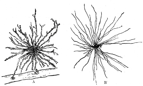
Fig. 623. Neuroglia cells of brain shown by Golgi’s method
A. Cell with branched processes.
B. Spider cell with unbranched processes. (After Andriezen.)
Neuroglia - the peculiar ground substance in which are imbedded the true nervous constituents of the brain and medulla spinalis, consists of cells and fibers. Some of the cells are stellate in shape, with ill-defined cell body, and their fine processes become neuroglia fibers, which extend radially and unbranched (Fig. 623, B) among the nerve cells and fibers which they aid in supporting. Other cells give off fibers which branch repeatedly (Fig. 623, A). Some of the fibers start from the epithelial cells lining the ventricles of the brain and central canal of the medulla spinalis, and pass through the nervous tissue, branching repeatedly to end in slight enlargements on the pia mater. Thus, neuroglia is evidently a connective tissue in function but is not so in development; it is ectodermal in origin, whereas all connective tissues are mesodermal.
- Gray's Images: Development | Lymphatic | Neural | Vision | Hearing | Somatosensory | Integumentary | Respiratory | Gastrointestinal | Urogenital | Endocrine | Surface Anatomy | iBook | Historic Disclaimer
| Historic Disclaimer - information about historic embryology pages |
|---|
| Pages where the terms "Historic" (textbooks, papers, people, recommendations) appear on this site, and sections within pages where this disclaimer appears, indicate that the content and scientific understanding are specific to the time of publication. This means that while some scientific descriptions are still accurate, the terminology and interpretation of the developmental mechanisms reflect the understanding at the time of original publication and those of the preceding periods, these terms, interpretations and recommendations may not reflect our current scientific understanding. (More? Embryology History | Historic Embryology Papers) |
| iBook - Gray's Embryology | |
|---|---|

|
|
Reference
Gray H. Anatomy of the human body. (1918) Philadelphia: Lea & Febiger.
Cite this page: Hill, M.A. (2024, April 16) Embryology Gray0623.jpg. Retrieved from https://embryology.med.unsw.edu.au/embryology/index.php/File:Gray0623.jpg
- © Dr Mark Hill 2024, UNSW Embryology ISBN: 978 0 7334 2609 4 - UNSW CRICOS Provider Code No. 00098G
File history
Click on a date/time to view the file as it appeared at that time.
| Date/Time | Thumbnail | Dimensions | User | Comment | |
|---|---|---|---|---|---|
| current | 22:33, 13 May 2013 |  | 500 × 301 (53 KB) | Z8600021 (talk | contribs) | {{Gray Anatomy}} |
You cannot overwrite this file.
File usage
The following page uses this file:

