File:Gray0621.jpg
Original file (599 × 800 pixels, file size: 131 KB, MIME type: image/jpeg)
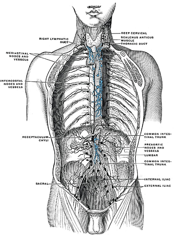
Thorax and Abdomen Deep Lymph Nodes and Vessels
(diagrammatic). Afferent vessels are represented by continuous lines, and efferent and internodular vessels by dotted lines. (Cunningham.)
The deep lymphatic vessels of the thoracic wall consist of:
- The lymphatics of the muscles which lie on the ribs: most of these end in the axillary glands, but some from the Pectoralis major pass to the sternal glands.
- The intercostal vessels which drain the Intercostales and parietal pleura. Those draining the Intercostales externi run backward and, after receiving the vessels which accompany the posterior branches of the intercostal arteries, end in the intercostal glands. Those of the Intercostales interni and parietal pleura consist of a single trunk in each space. These trunks run forward in the subpleural tissue and the upper six open separately into the sternal glands or into the vessels which unite them; those of the lower spaces unite to form a single trunk which terminates in the lowest of the sternal glands.
- The lymphatic vessels of the diaphragm, which form two plexuses, one on its thoracic and another on its abdominal surface. These plexuses anastomose freely with each other, and are best marked on the parts covered respectively by the pleuræ and peritoneum. That on the thoracic surface communicates with the lymphatics of the costal and mediastinal parts of the pleura, and its efferents consist of three groups: (a) anterior, passing to the gland which lie near the junction of the seventh rib with its cartilage; (b) middle, to the glands on the esophagus and to those around the termination of the inferior vena cava; and (c) posterior, to the glands which surround the aorta at the point where this vessel leaves the thoracic cavity.
The plexus on the abdominal surface is composed of fine vessels, and anastomoses with the lymphatics of the liver and, at the periphery of the diaphragm, with those of the subperitoneal tissue. The efferents from the right half of this plexus terminate partly in a group of glands on the trunk of the corresponding inferior phrenic artery, while others end in the right lateral aortic glands. Those from the left half of the plexus pass to the pre- and lateral aortic glands and to the glands on the terminal portion of the esophagus.
(Text from Gray's Anatomy 1918)
Gray's Lymphatic Anatomy: 592 Primary lymph sacs | 593 Lymph capillaries of the human conjunctiva | 594 Lymph capillaries from the human scrotum | 595 Lymph capillaries of the sole of the human foot | 596 Section through human tongue | 597 Lymph gland (Node) | 598 Lymph gland tissue | 599 Thoracic and right lymphatic ducts | 600 Modes of origin of thoracic duct | 601 Terminal collecting trunks of right side | 602 Lymph glands of the head | 603 Lymphatics of pharynx | 604 Lymphatics of the face | 605 Lymphatics of the Tongue | 606 Lymph glands of the upper extremity | 607 Lymphatics of the mamma | 608 Lymphatic vessels of the dorsal hand surface | 609 Lymph glands of popliteal fossa | 610 Superficial lymph glands and vessels of the lower extremity | 611 Parietal lymph glands of the pelvis | 612 Iliopelvic glands | 613 Lymphatics of stomach | 614 Lymphatics of stomach | 615 Lymphatics of cecum and vermiform process | 616 Lymphatics of cecum and vermiform process | 617 Lymphatics of Colon | 618 Lymphatic of the Bladder | 619 Lymphatics of the Prostate | 620 Lymphatics of the Uterus | 621 Lymphatics of the thorax and abdomen | 622 Tracheobronchial Lymph Glands | Gray's Anatomy | Historic Disclaimer | Lymphatic Development
- Gray's Images: Development | Lymphatic | Neural | Vision | Hearing | Somatosensory | Integumentary | Respiratory | Gastrointestinal | Urogenital | Endocrine | Surface Anatomy | iBook | Historic Disclaimer
| Historic Disclaimer - information about historic embryology pages |
|---|
 |
| iBook - Gray's Embryology | |
|---|---|

|
|
Reference
Gray H. Anatomy of the human body. (1918) Philadelphia: Lea & Febiger.
Cite this page: Hill, M.A. (2024, April 19) Embryology Gray0621.jpg. Retrieved from https://embryology.med.unsw.edu.au/embryology/index.php/File:Gray0621.jpg
- © Dr Mark Hill 2024, UNSW Embryology ISBN: 978 0 7334 2609 4 - UNSW CRICOS Provider Code No. 00098G
File history
Click on a date/time to view the file as it appeared at that time.
| Date/Time | Thumbnail | Dimensions | User | Comment | |
|---|---|---|---|---|---|
| current | 00:26, 15 February 2013 | | 599 × 800 (131 KB) | Z8600021 (talk | contribs) | ==Deep lymph nodes and vessels of the thorax and abdomen== (diagrammatic). Afferent vessels are represented by continuous lines, and efferent and internodular vessels by dotted lines. (Cunningham.) The deep lymphatic vessels of the thoracic wall consis |
You cannot overwrite this file.
File usage
The following 3 pages use this file: