File:Gray0607.jpg
Original file (800 × 623 pixels, file size: 126 KB, MIME type: image/jpeg)
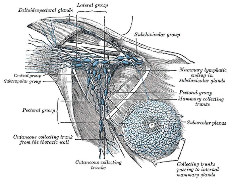
Lymphatics of the Mamma
Lymphatics of the mamma, and the axillary glands (semidiagrammatic). (Poirier and Charpy.)
The lymph glands of the upper extremity are divided into two sets, superficial and deep.
The deep lymph glands are chiefly grouped in the axilla, although a few may be found in the forearm, in the course of the radial, ulnar, and interosseous vessels, and in the arm along the medial side of the brachial artery. The Axillary Glands (lymphoglandulæ axillares) (Fig. 607) are of large size, vary from twenty to thirty in number, and may be arranged in the following groups:
- A lateral group of from four to six glands lies in relation to the medial and posterior aspects of the axillary vein; the afferents of these glands drain the whole arm with the exception of that portion whose vessels accompany the cephalic vein. The efferent vessels pass partly to the central and subclavicular groups of axillary glands and partly to the inferior deep cervical glands.
- An anterior or pectoral group consists of four or five glands along the lower border of the Pectoralis minor, in relation with the lateral thoracic artery. Their afferents drain the skin and muscles of the anterior and lateral thoracic walls, and the central and lateral parts of the namma; their efferents pass partly to the central and partly to the subclavicular groups of axillary glands.
- A posterior or subscapular group of six or seven glands is placed along the lower margin of the posterior wall of the axilla in the course of the subscapular artery. The afferents of this group drain the skin and muscles of the lower part of the back of the neck and of the posterior thoracic wall; their efferents pass to the central group of axillary glands.
- A central or intermediate group of three or four large glands is imbedded in the adipose tissue near the base of the axilla. Its afferents are the efferent vessels of all the preceding groups of axillary glands; its efferents pass to the subclavicular group.
- A medial or subclavicular group of six to twelve glands is situated partly posterior to the upper portion of the Pectoralis minor and partly above the upper border of this muscle. Its only direct territorial afferents are those which accompany the cephalic vein and one which drains the upper peripheral part of the mamma, but it receives the efferents of all the other axillary glands. The efferent vessels of the subclavicular group unite to form the subclavian trunk, which opens either directly into the junction of the internal jugular and subclavian veins or into the jugular lymphatic trunk; on the left side it may end in the thoracic duct. A few efferents from the subclavicular glands usually pass to the inferior deep cervical glands.
(Text from Gray's Anatomy 1918)
Gray's Lymphatic Anatomy: 592 Primary lymph sacs | 593 Lymph capillaries of the human conjunctiva | 594 Lymph capillaries from the human scrotum | 595 Lymph capillaries of the sole of the human foot | 596 Section through human tongue | 597 Lymph gland (Node) | 598 Lymph gland tissue | 599 Thoracic and right lymphatic ducts | 600 Modes of origin of thoracic duct | 601 Terminal collecting trunks of right side | 602 Lymph glands of the head | 603 Lymphatics of pharynx | 604 Lymphatics of the face | 605 Lymphatics of the Tongue | 606 Lymph glands of the upper extremity | 607 Lymphatics of the mamma | 608 Lymphatic vessels of the dorsal hand surface | 609 Lymph glands of popliteal fossa | 610 Superficial lymph glands and vessels of the lower extremity | 611 Parietal lymph glands of the pelvis | 612 Iliopelvic glands | 613 Lymphatics of stomach | 614 Lymphatics of stomach | 615 Lymphatics of cecum and vermiform process | 616 Lymphatics of cecum and vermiform process | 617 Lymphatics of Colon | 618 Lymphatic of the Bladder | 619 Lymphatics of the Prostate | 620 Lymphatics of the Uterus | 621 Lymphatics of the thorax and abdomen | 622 Tracheobronchial Lymph Glands | Gray's Anatomy | Historic Disclaimer | Lymphatic Development
- Gray's Images: Development | Lymphatic | Neural | Vision | Hearing | Somatosensory | Integumentary | Respiratory | Gastrointestinal | Urogenital | Endocrine | Surface Anatomy | iBook | Historic Disclaimer
| Historic Disclaimer - information about historic embryology pages |
|---|
 |
| iBook - Gray's Embryology | |
|---|---|

|
|
Reference
Gray H. Anatomy of the human body. (1918) Philadelphia: Lea & Febiger.
Cite this page: Hill, M.A. (2024, April 19) Embryology Gray0607.jpg. Retrieved from https://embryology.med.unsw.edu.au/embryology/index.php/File:Gray0607.jpg
- © Dr Mark Hill 2024, UNSW Embryology ISBN: 978 0 7334 2609 4 - UNSW CRICOS Provider Code No. 00098G
File history
Click on a date/time to view the file as it appeared at that time.
| Date/Time | Thumbnail | Dimensions | User | Comment | |
|---|---|---|---|---|---|
| current | 16:18, 14 February 2013 | | 800 × 623 (126 KB) | Z8600021 (talk | contribs) | ==Lymphatics of the Mamma== Lymphatics of the mamma, and the axillary glands (semidiagrammatic). (Poirier and Charpy.) The lymph glands of the upper extremity are divided into two sets, superficial and deep. The deep lymph glands are chiefly grouped in |
You cannot overwrite this file.
File usage
The following 3 pages use this file: