File:Gray0589.jpg

Original file (900 × 534 pixels, file size: 134 KB, MIME type: image/jpeg)
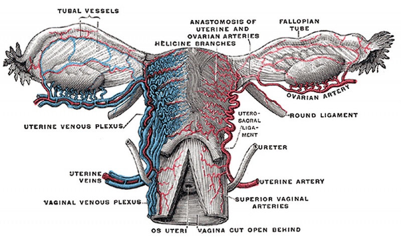
Fig. 589. Vessels of the uterus and its appendages
Rear view. (Testut.)
Uterine Plexuses
Lie along the sides and superior angles of the uterus between the two layers of the broad ligament, and communicate with the ovarian and vaginal plexuses. They are drained by a pair of uterine veins on either side: these arise from the lower part of the plexuses, opposite the external orifice of the uterus, and open into the corresponding hypogastric vein.
Vaginal Plexuses
Placed at the sides of the vagina; they communicate with the uterine, vesical, and hemorrhoidal plexuses, and are drained by the vaginal veins, one on either side, into the hypogastric veins.
Ovarian Veins
(vv. ovaricæ) correspond with the spermatic in the male; they form a plexus in the broad ligament near the ovary and uterine tube, and communicate with the uterine plexus. They end in the same way as the spermatic veins in the male. Valves are occasionally found in these veins. Like the uterine veins, they become much enlarged during pregnancy.
- Gray's Images: Development | Lymphatic | Neural | Vision | Hearing | Somatosensory | Integumentary | Respiratory | Gastrointestinal | Urogenital | Endocrine | Surface Anatomy | iBook | Historic Disclaimer
| Historic Disclaimer - information about historic embryology pages |
|---|
 |
| iBook - Gray's Embryology | |
|---|---|

|
|
Reference
Gray H. Anatomy of the human body. (1918) Philadelphia: Lea & Febiger.
Cite this page: Hill, M.A. (2024, April 19) Embryology Gray0589.jpg. Retrieved from https://embryology.med.unsw.edu.au/embryology/index.php/File:Gray0589.jpg
- © Dr Mark Hill 2024, UNSW Embryology ISBN: 978 0 7334 2609 4 - UNSW CRICOS Provider Code No. 00098G
File history
Click on a date/time to view the file as it appeared at that time.
| Date/Time | Thumbnail | Dimensions | User | Comment | |
|---|---|---|---|---|---|
| current | 16:05, 9 June 2013 | | 900 × 534 (134 KB) | Z8600021 (talk | contribs) | ==Fig. 589. Vessels of the uterus and its appendages== Rear view. (Testut.) ===Uterine plexuses=== Lie along the sides and superior angles of the uterus between the two layers of the broad ligament, and communicate with the ovarian and vaginal plex... |
You cannot overwrite this file.
File usage
The following 3 pages use this file: