File:Gray0502.jpg
Original file (1,000 × 1,329 pixels, file size: 215 KB, MIME type: image/jpeg)
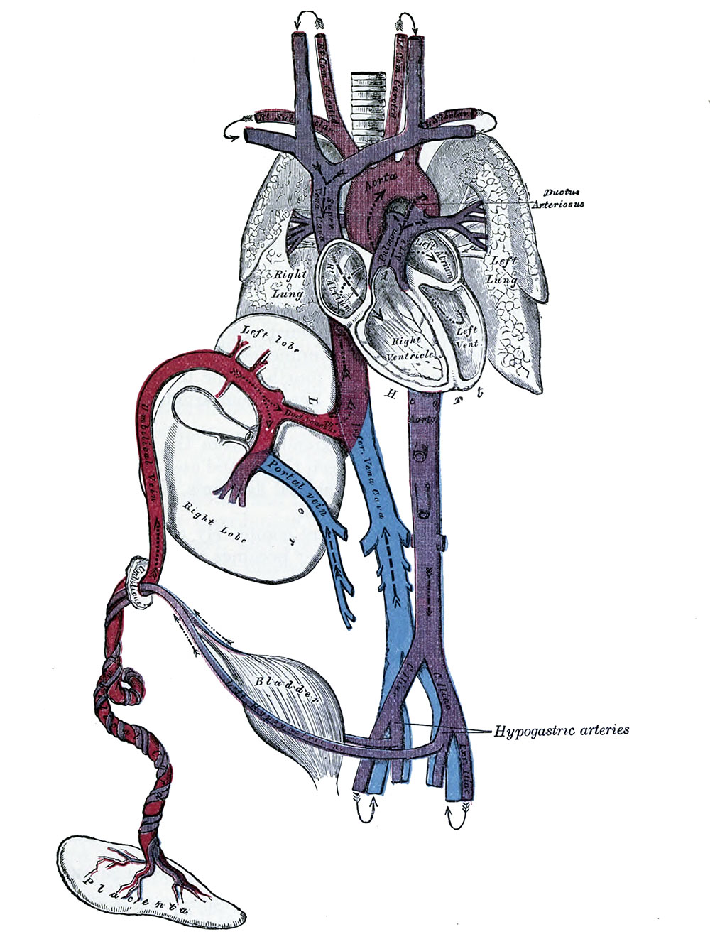
Fetal Circulation
Plan of the fetal circulation. In this plan the figured arrows represent the kind of blood, as well as the direction which it takes in the vessels.
Thus—arterial blood is figured >—>; venous blood, >--->; mixed (arterial and venous) blood, >—>.
The fetal blood is returned from the placenta to the fetus by the umbilical vein. This vein enters the abdomen at the umbilicus, and passes upward along the free margin of the falciform ligament of the liver to the under surface of that organ, where it gives off two or three branches, one of large size to the left lobe, and others to the lobus quadratus and lobus caudatus. At the porta hepatis (transverse fissure of the liver) it divides into two branches: of these, the larger is joined by the portal vein, and enters the right lobe; the smaller is continued upward, under the name of the ductus venosus, and joins the inferior vena cava. The blood, therefore, which traverses the umbilical vein, passes to the inferior vena cava in three different ways. A considerable quantity circulates through the liver with the portal venous blood, before entering the inferior vena cava by the hepatic veins; some enters the liver directly, and is carried to the inferior cava by the hepatic veins; the remainder passes directly into the inferior vena cava through the ductus venous.
In the inferior vena cava, the blood carried by the ductus venosus and hepatic veins becomes mixed with that returning from the lower extremities and abdominal wall. It enters the right atrium, and, guided by the valve of the inferior vena cava, passes through the formen ovale into the left atrium, where it mixes with a small quantity of blood returned from the lungs by the pulmonary veins. From the left atrium it passes into the left ventricle; and from the left ventricle into the aorta, by means of which it is distributed almost entirely to the head and upper extremities, a small quantity being probably carried into the descending aorta. From the head and upper extremities the blood is returned by the superior vena cava to the right atrium, where it mixes with a small portion of the blood from the inferior vena cava. From the right atrium it descends into the right ventricle, and thence passes into the pulmonary artery. The lungs of the fetus being inactive, only a small quantity of the blood of the pulmonary artery is distributed to them by the right and left pulmonary arteries, and returned by the pulmonary veins to the left atrium: the greater part passes through the ductus arteriosus into the aorta, where it mixes with a small quantity of the blood transmitted by the left ventricle into the aorta. Through this vessel it descends, and is in part distributed to the lower extremities and the viscera of the abdomen and pelvis, but the greater amount is conveyed by the umbilical arteries to the placenta.
From the preceding account of the circulation of the blood in the fetus the following facts will be evident:
- The placenta serves the purposes of nutrition and excretion, receiving the impure blood from the fetus, and returning it purified and charged with additional nutritive material.
- Nearly the whole of the blood of the umbilical vein traverses the liver before entering the inferior vena cava; hence the large size of the liver, especially at an early period of fetal life.
- The right atrium is the point of meeting of a double current, the blood in the inferior vena cava being guided by the valve of this vessel into the left atrium, while that in the superior vena cava descends into the right ventricle. At an early period of fetal life it is highly probable that the two streams are quite distinct; for the inferior vena cava opens almost directly into the left atrium, and the valve of the inferior vena cava would exclude the current from the right ventricle. At a later period, as the separation between the two atria becomes more distinct, it seems probable that some mixture of the two streams must take place.
- The pure blood carried from the placenta to the fetus by the umbilical vein, mixed with the blood from the portal vein and inferior vena cava, passes almost directly to the arch of the aorta, and is distributed by the branches of that vessel to the head and upper extremities.
- The blood contained in the descending aorta, chiefly derived from that which has already circulated through the head and limbs, together with a small quantity from the left ventricle, is distributed to the abdomen and lower extremities.
(Text modified from Gray's Anatomy)
- Gray's Images: Development | Lymphatic | Neural | Vision | Hearing | Somatosensory | Integumentary | Respiratory | Gastrointestinal | Urogenital | Endocrine | Surface Anatomy | iBook | Historic Disclaimer
| Historic Disclaimer - information about historic embryology pages |
|---|
 |
| iBook - Gray's Embryology | |
|---|---|

|
|
Reference
Gray H. Anatomy of the human body. (1918) Philadelphia: Lea & Febiger.
Cite this page: Hill, M.A. (2024, April 19) Embryology Gray0502.jpg. Retrieved from https://embryology.med.unsw.edu.au/embryology/index.php/File:Gray0502.jpg
- © Dr Mark Hill 2024, UNSW Embryology ISBN: 978 0 7334 2609 4 - UNSW CRICOS Provider Code No. 00098G
File history
Click on a date/time to view the file as it appeared at that time.
| Date/Time | Thumbnail | Dimensions | User | Comment | |
|---|---|---|---|---|---|
| current | 21:34, 9 June 2014 | | 1,000 × 1,329 (215 KB) | Z8600021 (talk | contribs) | |
| 18:08, 30 May 2012 |  | 620 × 1,000 (103 KB) | Z8600021 (talk | contribs) | ==Fetal Circulation== Plan of the fetal circulation. In this plan the figured arrows represent the kind of blood, as well as the direction which it takes in the vessels. Thus—arterial blood is figured >—>; venous blood, >--->; mixed (arterial and v |
You cannot overwrite this file.
File usage
The following 7 pages use this file: