File:Gray0480.jpg
Gray0480.jpg (597 × 560 pixels, file size: 31 KB, MIME type: image/jpeg)
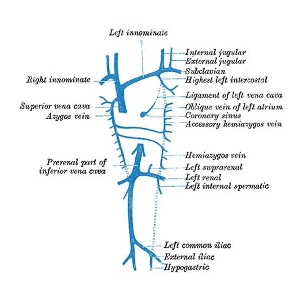
Diagram showing development of main cross branches between jugulars and between cardinals
Above the level of the renal veins the right cardinal vein persists as the azygos vein and receives the right intercostal veins, while the hemiazygos veins are brought into communication with it by the development of transverse branches in front of the vertebral column (Figs. 479, 480)
The portion of the right subcardinal behind this cross communication disappears, while that in front, i.e., the prerenal part, forms a connection with the ductus venosus at the point of opening of the hepatic veins, and, rapidly enlarging, receives the blood from the postrenal part of the right cardinal through the cross communication referred to. In this manner a single trunk, the inferior vena cava (Fig. 480), is formed, and consists of the proximal part of the ductus venosus, the prerenal part of the right subcardinal vein, the postrenal part of the right cardinal vein, and the cross branch which joins these two veins.
The right and left Cuvierian ducts are originally of the same diameter, and are frequently termed the right and left superior venæ cavæ. By the development of a transverse branch, the left innominate vein between the two primitive jugular veins, the blood is carried across from the left to the right primitive jugular (Figs. 479, 480). The portion of the right primitive jugular vein between the left innominate and the azygos vein forms the upper part of the superior vena cava of the adult; the lower part of this vessel, i.e., below the entrance of the azygos vein, is formed by the right Cuvierian duct. Below the origin of the transverse branch the left primitive jugular vein and left Cuvierian duct atrophy, the former constituting the upper part of the highest left intercostal vein, while the latter is represented by the ligament of the left vena cava, vestigial fold of Marshall, and the oblique vein of the left atrium, oblique vein of Marshall (Fig. 480). Both right and left superior venæ cavæ are present in some animals, and are occasionally found in the adult human being. The oblique vein of the left atrium passes downward across the back of the left atrium to open into the coronary sinus, which, as already indicated, represents the persistent left horn of the sinus venosus.
- Venous Links: early parietal veins | inferior vena cava | jugular and cardinal cross branches | completed parietal veins
- Gray's Images: Development | Lymphatic | Neural | Vision | Hearing | Somatosensory | Integumentary | Respiratory | Gastrointestinal | Urogenital | Endocrine | Surface Anatomy | iBook | Historic Disclaimer
| Historic Disclaimer - information about historic embryology pages |
|---|
| Pages where the terms "Historic" (textbooks, papers, people, recommendations) appear on this site, and sections within pages where this disclaimer appears, indicate that the content and scientific understanding are specific to the time of publication. This means that while some scientific descriptions are still accurate, the terminology and interpretation of the developmental mechanisms reflect the understanding at the time of original publication and those of the preceding periods, these terms, interpretations and recommendations may not reflect our current scientific understanding. (More? Embryology History | Historic Embryology Papers) |
| iBook - Gray's Embryology | |
|---|---|

|
|
Reference
Gray H. Anatomy of the human body. (1918) Philadelphia: Lea & Febiger.
Cite this page: Hill, M.A. (2024, April 19) Embryology Gray0480.jpg. Retrieved from https://embryology.med.unsw.edu.au/embryology/index.php/File:Gray0480.jpg
- © Dr Mark Hill 2024, UNSW Embryology ISBN: 978 0 7334 2609 4 - UNSW CRICOS Provider Code No. 00098G
File history
Click on a date/time to view the file as it appeared at that time.
| Date/Time | Thumbnail | Dimensions | User | Comment | |
|---|---|---|---|---|---|
| current | 23:18, 11 October 2009 |  | 597 × 560 (31 KB) | S8600021 (talk | contribs) |
You cannot overwrite this file.

