File:Gray0448.jpg
Gray0448.jpg (356 × 600 pixels, file size: 70 KB, MIME type: image/jpeg)
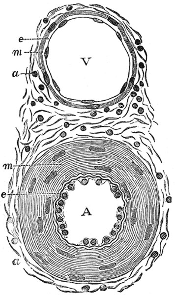
Fig. 448 Transverse section through a small artery and vein
Transverse section through a small artery and vein of the mucous membrane of the epiglottis of a child. X 350. (Klein and Noble Smith.)
A. Artery - showing the nucleated endothelium, e, which lines it; the vessel being contracted, the endothelial cells appear very thick. Underneath the endothelium is the wavy elastic lamina. The chief part of the wall of the vessel is occupied by the circular muscle coat m; the rod-shaped nuclei of the muscle cells are well seen. Outside this is a, part of the adventitia. This is composed of bundles of connective tissue fibers, shown in section, with the nuclei of the connective tissue corpuscles. The adventitia gradually merges into the surrounding connective tissue.
V. Vein - showing a thin endothelial membrane, e, raised accidentally from the intima, which on account of its delicacy is seen as a mere line on the media m. This latter is composed of a few circular unstriped muscle cells a. The adventitia, similar in structure to that of an artery.
| Historic Disclaimer - information about historic embryology pages |
|---|
| Pages where the terms "Historic" (textbooks, papers, people, recommendations) appear on this site, and sections within pages where this disclaimer appears, indicate that the content and scientific understanding are specific to the time of publication. This means that while some scientific descriptions are still accurate, the terminology and interpretation of the developmental mechanisms reflect the understanding at the time of original publication and those of the preceding periods, these terms, interpretations and recommendations may not reflect our current scientific understanding. (More? Embryology History | Historic Embryology Papers) |
Historic Description of Artery Layers
tunica intima
The inner coat can be separated from the middle by a little maceration, or it may be stripped off in small pieces; but, on account of its friability, it cannot be separated as a complete membrane. It is a fine, transparent, colorless structure which is highly elastic, and, after death, is commonly corrugated into longitudinal wrinkles. The inner coat consists of:
- A layer of pavement endothelium, the cells of which are polygonal, oval, or fusiform, and have very distinct round or oval nuclei. This endothelium is brought into view most distinctly by staining with nitrate of silver.
- A subendothelial layer, consisting of delicate connective tissue with branched cells lying in the interspaces of the tissue; in arteries of less than 2 mm. in diameter the subendothelial layer consists of a single stratum of stellate cells, and the connective tissue is only largely developed in vessels of a considerable size.
- An elastic or fenestrated layer, which consists of a membrane containing a net-work of elastic fibers, having principally a longitudinal direction, and in which, under the microscope, small elongated apertures or perforations may be seen, giving it a fenestrated appearance. It was therefore called by Henle the fenestrated membrane. This membrane forms the chief thickness of the inner coat, and can be separated into several layers, some of which present the appearance of a net-work of longitudinal elastic fibers, and others a more membranous character, marked by pale lines having a longitudinal direction. In minute arteries the fenestrated membrane is a very thin layer; but in the larger arteries, and especially in the aorta, it has a very considerable thickness.
tunica media
The middle coat is distinguished from the inner by its color and by the transverse arrangement of its fibers. In the smaller arteries it consists principally of plain muscle fibers in fine bundles, arranged in lamellæ and disposed circularly around the vessel. These lamellæ vary in number according to the size of the vessel; the smallest arteries having only a single layer (Fig. 449), and those slightly larger three or four layers. It is to this coat that the thickness of the wall of the artery is mainly due (Fig. 448 A, m). In the larger arteries, as the iliac, femoral, and carotid, elastic fibers unite to form lamellæ which alternate with the layers of muscular fibers; these lamellæ are united to one another by elastic fibers which pass between the muscular bundles, and are connected with the fenestrated membrane of the inner coat (Fig. 450). In the largest arteries, as the aorta and innominate, the amount of elastic tissue is very considerable; in these vessels a few bundles of white connective tissue also have been found in the middle coat. The muscle fiber cells are about 50μ in length and contain well-marked, rod-shaped nuclei, which are often slightly curved.
tunica adventitia
The external coat consists mainly of fine and closely felted bundles of white connective tissue, but also contains elastic fibers in all but the smallest arteries. The elastic tissue is much more abundant next the tunica media, and it is sometimes described as forming here, between the adventitia and media, a special layer, the tunica elastica externa of Henle. This layer is most marked in arteries of medium size. In the largest vessels the external coat is relatively thin; but in small arteries it is of greater proportionate thickness. In the smaller arteries it consists of a single layer of white connective tissue and elastic fibers; while in the smallest arteries, just above the capillaries, the elastic fibers are wanting, and the connective tissue of which the coat is composed becomes more nearly homogeneous the nearer it approaches the capillaries, and is gradually reduced to a thin membranous envelope, which finally disappears.
- Gray - Cardiovascular: 448 Artery and vein | 458 Human embryo week 2 vascular | 459 Human embryo 14 days with yolk-sac
- Gray's Images: Development | Lymphatic | Neural | Vision | Hearing | Somatosensory | Integumentary | Respiratory | Gastrointestinal | Urogenital | Endocrine | Surface Anatomy | iBook | Historic Disclaimer
| Historic Disclaimer - information about historic embryology pages |
|---|
| Pages where the terms "Historic" (textbooks, papers, people, recommendations) appear on this site, and sections within pages where this disclaimer appears, indicate that the content and scientific understanding are specific to the time of publication. This means that while some scientific descriptions are still accurate, the terminology and interpretation of the developmental mechanisms reflect the understanding at the time of original publication and those of the preceding periods, these terms, interpretations and recommendations may not reflect our current scientific understanding. (More? Embryology History | Historic Embryology Papers) |
| iBook - Gray's Embryology | |
|---|---|

|
|
Reference
Gray H. Anatomy of the human body. (1918) Philadelphia: Lea & Febiger.
Cite this page: Hill, M.A. (2024, April 19) Embryology Gray0448.jpg. Retrieved from https://embryology.med.unsw.edu.au/embryology/index.php/File:Gray0448.jpg
- © Dr Mark Hill 2024, UNSW Embryology ISBN: 978 0 7334 2609 4 - UNSW CRICOS Provider Code No. 00098G
File history
Click on a date/time to view the file as it appeared at that time.
| Date/Time | Thumbnail | Dimensions | User | Comment | |
|---|---|---|---|---|---|
| current | 14:08, 6 July 2012 |  | 356 × 600 (70 KB) | Z8600021 (talk | contribs) | FIG. 448– Transverse section through a small artery and vein of the mucous membrane of the epiglottis of a child. X 350. (Klein and Noble Smith.) A. Artery, showing the nucleated endothelium, e, which lines it; the vessel being contracted, the endothel |
You cannot overwrite this file.
File usage
The following 2 pages use this file:

