File:Gray0321.jpg
Original file (800 × 611 pixels, file size: 56 KB, MIME type: image/jpeg)
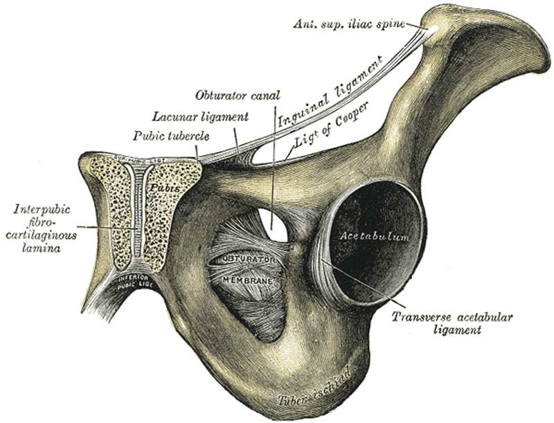
Fig. 321. Symphysis Pubis exposed by a Coronal Section
The Pubic Symphysis (symphysis ossium pubis; articulation of the pubic bones) (Fig. 321 The articulation between the pubic bones is an amphiarthrodial joint, formed between the two oval articular surfaces of the bones.
The ligaments of this articulation are: Anterior Pubic, Posterior Pubic, Superior Pubic, Arcuate Pubic and Interpubic Fibrocartilaginous Lamina.
Anterior Pubic Ligament
(Fig. 319) The anterior pubic ligament consists of several superimposed layers, which pass across the front of the articulation. The superficial fibers pass obliquely from one bone to the other, decussating and forming an interlacement with the fibers of the aponeuroses of the Obliqui externi and the medial tendons of origin of the Recti abdominis. The deep fibers pass transversely across the symphysis, and are blended with the fibrocartilaginous lamina.
Posterior Pubic Ligament
The posterior pubic ligament consists of a few thin, scattered fibers, which unite the two pubic bones posteriorly.
Superior Pubic Ligament
(ligamentum pubicum superius) The superior pubic ligament connects together the two pubic bones superiorly, extending laterally as far as the pubic tubercles.
Arcuate Pubic Ligament
(ligamentum arcuatum pubis; inferior pubic or subpubic ligament) The arcuate pubic ligament is a thick, triangular arch of ligamentous fibers, connecting together the two pubic bones below, and forming the upper boundary of the pubic arch. Above, it is blended with the interpubic fibrocartilaginous lamina; laterally, it is attached to the inferior rami of the pubic bones; below, it is free, and is separated from the fascia of the urogenital diaphragm by an opening through which the deep dorsal vein of the penis passes into the pelvis.
Interpubic Fibrocartilaginous Lamina
(lamina fibrocartilaginea interpubica; interpubic disk) The interpubic fibrocartilaginous lamina connects the opposed surfaces of the pubic bones. Each of these surfaces is covered by a thin layer of hyaline cartilage firmly joined to the bone by a series of nipple-like processes which accurately fit into corresponding depressions on the osseous surfaces. These opposed cartilaginous surfaces are connected together by an intermediate lamina of fibrocartilage which varies in thickness in different subjects. It often contains a cavity in its interior, probably formed by the softening and absorption of the fibrocartilage, since it rarely appears before the tenth year of life and is not lined by synovial membrane. This cavity is larger in the female than in the male, but it is very doubtful whether it enlarges, as was formerly supposed, during pregnancy. It is most frequently limited to the upper and back part of the joint; it occasionally reaches to the front, and may extend the entire length of the cartilage. It may be easily demonstrated when present by making a coronal section of the symphysis pubis near its posterior surface.
- Gray's Images: Development | Lymphatic | Neural | Vision | Hearing | Somatosensory | Integumentary | Respiratory | Gastrointestinal | Urogenital | Endocrine | Surface Anatomy | iBook | Historic Disclaimer
| Historic Disclaimer - information about historic embryology pages |
|---|
 |
| iBook - Gray's Embryology | |
|---|---|

|
|
Reference
Gray H. Anatomy of the human body. (1918) Philadelphia: Lea & Febiger.
Cite this page: Hill, M.A. (2024, April 25) Embryology Gray0321.jpg. Retrieved from https://embryology.med.unsw.edu.au/embryology/index.php/File:Gray0321.jpg
- © Dr Mark Hill 2024, UNSW Embryology ISBN: 978 0 7334 2609 4 - UNSW CRICOS Provider Code No. 00098G
File history
Click on a date/time to view the file as it appeared at that time.
| Date/Time | Thumbnail | Dimensions | User | Comment | |
|---|---|---|---|---|---|
| current | 13:03, 4 January 2011 | | 800 × 611 (56 KB) | S8600021 (talk | contribs) | {{Gray Anatomy}} |
You cannot overwrite this file.
File usage
The following 6 pages use this file: