File:Gray0054.jpg
Gray0054.jpg (800 × 513 pixels, file size: 71 KB, MIME type: image/jpeg)
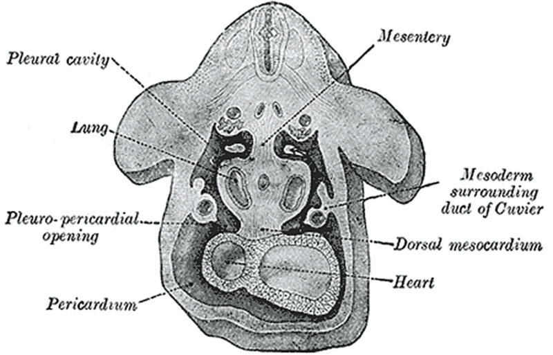
Week 4 - Coelomic Cavity
Figure obtained by combining several successive sections of a human embryo of about the fourth week (From Kollmann.) The upper arrow is in the pleuroperitoneal opening, the lower in the pleuropericardial.
In the human embryo described by Peters the mesoderm outside the embryonic disk is split into two layers enclosing an extra-embryonic cœlom; there is no trace of an intra-embryonic cœlom. At a later stage four cavities are formed within the embryo, viz., one on either side within the mesoderm of the pericardial area, and one in either lateral mass of the general mesoderm. All these are at first independent of each other and of the extra-embryonic celom, but later they become continuous. The two cavities in the general mesoderm unite on the ventral aspect of the gut and form the pleuro-peritoneal cavity, which becomes continuous with the remains of the extra-embryonic celom around the umbilicus; the two cavities in the pericardial area rapidly join to form a single pericardial cavity, and this from two lateral diverticula extend caudalward to open into the pleuro-peritoneal cavity
- Gray's Images: Development | Lymphatic | Neural | Vision | Hearing | Somatosensory | Integumentary | Respiratory | Gastrointestinal | Urogenital | Endocrine | Surface Anatomy | iBook | Historic Disclaimer
| Historic Disclaimer - information about historic embryology pages |
|---|
 |
| iBook - Gray's Embryology | |
|---|---|

|
|
Reference
Gray H. Anatomy of the human body. (1918) Philadelphia: Lea & Febiger.
Cite this page: Hill, M.A. (2024, April 25) Embryology Gray0054.jpg. Retrieved from https://embryology.med.unsw.edu.au/embryology/index.php/File:Gray0054.jpg
- © Dr Mark Hill 2024, UNSW Embryology ISBN: 978 0 7334 2609 4 - UNSW CRICOS Provider Code No. 00098G
File history
Click on a date/time to view the file as it appeared at that time.
| Date/Time | Thumbnail | Dimensions | User | Comment | |
|---|---|---|---|---|---|
| current | 17:32, 28 April 2011 | | 800 × 513 (71 KB) | S8600021 (talk | contribs) | ==Week 4 - Coelomic Cavity== Figure obtained by combining several successive sections of a human embryo of about the fourth week (From Kollmann.) The upper arrow is in the pleuroperitoneal opening, the lower in the pleuropericardial. {{Gray Anatomy}} [ |
You cannot overwrite this file.
File usage
There are no pages that use this file.