File:Gray0043.jpg
Gray0043.jpg (800 × 496 pixels, file size: 50 KB, MIME type: image/jpeg)
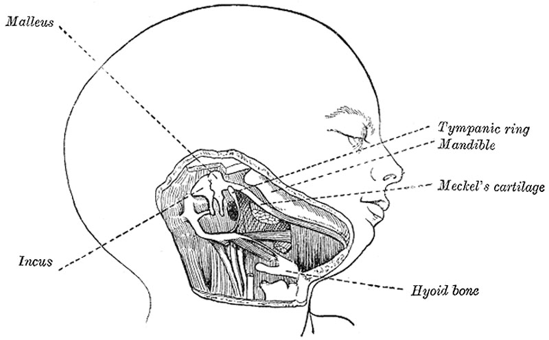
Head and neck of a human embryo eighteen weeks old with Meckel’s cartilage and hyoid bar exposed
(After Kölliker.)
The mandibular arch lies between the first branchial groove and the stomodeum; from it are developed the lower lip, the mandible, the muscles of mastication, and the anterior part of the tongue. Its cartilaginous bar is formed by what are known as Meckel’s cartilages (right and left) (Fig. 43); above this the incus is developed. The dorsal end of each cartilage is connected with the ear-capsule and is ossified to form the malleus; the ventral ends meet each other in the region of the symphysis menti, and are usually regarded as undergoing ossification to form that portion of the mandible which contains the incisor teeth. The intervening part of the cartilage disappears; the portion immediately adjacent to the malleus is replaced by fibrous membrane, which constitutes the spheno-mandibular ligament, while from the connective tissue covering the remainder of the cartilage the greater part of the mandible is ossified.
- Gray's Images: Development | Lymphatic | Neural | Vision | Hearing | Somatosensory | Integumentary | Respiratory | Gastrointestinal | Urogenital | Endocrine | Surface Anatomy | iBook | Historic Disclaimer
| Historic Disclaimer - information about historic embryology pages |
|---|
 |
| iBook - Gray's Embryology | |
|---|---|

|
|
Reference
Gray H. Anatomy of the human body. (1918) Philadelphia: Lea & Febiger.
Cite this page: Hill, M.A. (2024, April 23) Embryology Gray0043.jpg. Retrieved from https://embryology.med.unsw.edu.au/embryology/index.php/File:Gray0043.jpg
- © Dr Mark Hill 2024, UNSW Embryology ISBN: 978 0 7334 2609 4 - UNSW CRICOS Provider Code No. 00098G
File history
Click on a date/time to view the file as it appeared at that time.
| Date/Time | Thumbnail | Dimensions | User | Comment | |
|---|---|---|---|---|---|
| current | 14:38, 22 April 2013 | | 800 × 496 (50 KB) | Z8600021 (talk | contribs) | ==Head and neck of a human embryo eighteen weeks old with Meckel’s cartilage and hyoid bar exposed== (After Kölliker.) {{Gray Anatomy}} Category:Head Category:Hearing Category:Cartoon |
You cannot overwrite this file.
File usage
The following page uses this file: