File:Gland duct histology cartoon.jpg
Gland_duct_histology_cartoon.jpg (600 × 400 pixels, file size: 41 KB, MIME type: image/jpeg)
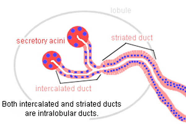
Gland Duct Histology Cartoon
Cartoon shows the general organisation of salivary gland duct system.
Interlobular and interlobar ducts - embedded in the connective tissue surrounding the lobes and lobules of the glands
stratified cuboidal or stratified columnar epithelium
stratified squamous epithelium at the oral cavity opening
Intralobular ducts - in between the secretory acini within the lobules
Intercalated ducts - difficult to identify in mucous glands
Striated ducts - absent in purely mucous glands
- Parotid Gland Links: Gland overview | lobe overview | serous acini | Striated duct and serous | Intercalated duct | excretory duct | Gland epithelium | Duct cartoon
Cite this page: Hill, M.A. (2024, April 25) Embryology Gland duct histology cartoon.jpg. Retrieved from https://embryology.med.unsw.edu.au/embryology/index.php/File:Gland_duct_histology_cartoon.jpg
- © Dr Mark Hill 2024, UNSW Embryology ISBN: 978 0 7334 2609 4 - UNSW CRICOS Provider Code No. 00098G
File history
Click on a date/time to view the file as it appeared at that time.
| Date/Time | Thumbnail | Dimensions | User | Comment | |
|---|---|---|---|---|---|
| current | 10:55, 26 March 2014 | | 600 × 400 (41 KB) | Z8600021 (talk | contribs) |
You cannot overwrite this file.
File usage
The following 3 pages use this file: