File:Germ cell tumor 01.jpg
Germ_cell_tumor_01.jpg (800 × 525 pixels, file size: 161 KB, MIME type: image/jpeg)
Teratoma - Germ Cell Tumor
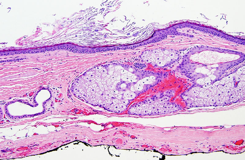
The image shows a teratoma located within the lesser sac of the momentum (see pathology description below).
Germ cell tumours (germ cell tumors, GCT, teratomas) are a neoplasm derived from primordial germ cells and occur in the gonads, but may rarely be found outside of the gonads (extra-gonadal) mainly in midline structures. Primary abdominal or retroperitoneal germ cell tumors comprise approximately 5% of extragonadal tumours.
Pathologic examination revealed an 8.5 × 7.9 × 7.0 cm cystic structure, which on sectioning demonstrated grumose (clustered in grains at intervals) material with a moderate amount of hair. No teeth or calcified structures were identified. On microscopic examination, the cyst was lined by stratified squamous epithelium with associated hair follicles, sebaceous glands, and sweat glands.
- Links: teratoma 1 | teratoma 2 | Testis Development
Reference
<pubmed>22606456</pubmed>| Case Rep Oncol Med.
Copyright © 2012 Brandon M. Hardesty et al. This is an open access article distributed under the Creative Commons Attribution License, which permits unrestricted use, distribution, and reproduction in any medium, provided the original work is properly cited.
604571.fig.002.jpg
File history
Click on a date/time to view the file as it appeared at that time.
| Date/Time | Thumbnail | Dimensions | User | Comment | |
|---|---|---|---|---|---|
| current | 08:28, 29 May 2012 |  | 800 × 525 (161 KB) | Z8600021 (talk | contribs) | ==Germ Cell Tumor== The image shows a teratoma located within the lesser sac of the omentum. Germ cell tumors (tumours, GCT) are a neoplasm derived from primordial germ cells and occur in the gonads, but may rarely be found outside of the gonads (extra-g |
You cannot overwrite this file.
File usage
There are no pages that use this file.
