File:GemmillStewart1916 fig03.jpg

Original file (1,280 × 660 pixels, file size: 89 KB, MIME type: image/jpeg)
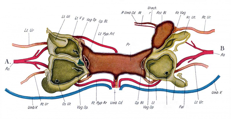
Fig. 3. Diagrammatic representation of a dissection of the urogenital system of the twins
Looked at from above. The cavities of the vagina: and bladders have been opened up so as to exhibit the floor of each with the various openings leading therefrom.
A, on side of twin with club-foot.
B, on side of twin without club-foot.
Note - Clubfoot is a historic term for talipes equinovarus.
| Abbreviations | ||
|---|---|---|
|
|
|
Reference
Gemmill JF. and Stewart J. Omphalopagous twins in the human subject. (1916) J Anat Physiol. 50: 316-323.
Cite this page: Hill, M.A. (2024, April 23) Embryology GemmillStewart1916 fig03.jpg. Retrieved from https://embryology.med.unsw.edu.au/embryology/index.php/File:GemmillStewart1916_fig03.jpg
- © Dr Mark Hill 2024, UNSW Embryology ISBN: 978 0 7334 2609 4 - UNSW CRICOS Provider Code No. 00098G
File history
Click on a date/time to view the file as it appeared at that time.
| Date/Time | Thumbnail | Dimensions | User | Comment | |
|---|---|---|---|---|---|
| current | 20:17, 15 March 2018 | | 1,280 × 660 (89 KB) | Z8600021 (talk | contribs) | |
| 20:11, 15 March 2018 |  | 2,289 × 1,380 (309 KB) | Z8600021 (talk | contribs) |
You cannot overwrite this file.
File usage
The following page uses this file: