File:Gage1905-plate4.jpg

Original file (2,194 × 1,500 pixels, file size: 450 KB, MIME type: image/jpeg)
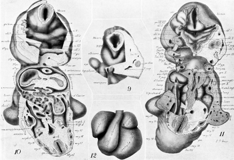
Plate IV
Fig. 9. A segment of the model from section 162 to section 200 (cf. Figs. 1-4), showing: A caudal view of the eye vesicle and visual lobe; the V-shaped union of the albicantial folds and their independent dorsal ending; the mesencephal with its sharpened beak-like ventral ending between the albicantial folds; the strand of tissue, at the point where the III N. would later appear; the hypophysis forming a bi-lobed, ectodermic organ surrounding the end of the hypophysial fold ; part of the neuroporic thickening; and the Gasserian ganglion.
Fig. 10. A segment of the model extending from Sec. 155, through the cephalic flexure, to Sec. 96. With dissections at Secs. 130 and 140. It shows: The base of the mesencephal and oblongata, with the large protuberance (4) at the end of the second total fold of the mesencephal; the oblongata folds 1-5, and the relations to the Vth and VIIth Ns.; mouth; pharynx; the ending of the gill-clefts 2-4 in the precervical sinus; the entrance of the vitelline veins into the liver at the side of the duodenum and their union in the dorsal part of the liver with the sinus venosus; the vitelline artery; and the mesonephros.
Fig. 11. A View from the dorsal side of the same segment of the model as is shown from the ventral side in Fig. 10, i. 9., it extends from See. 96 to Sec. 155 (of. Figs. 1, 3, 4). It cuts the arm buds. looks into the door of the pharynx and cephalad into the pons, mesencephal and ear vesicles.
There are seen: A portion of the cerebellum with its folds; the mesencephal with its narrow opening cephalad and its floor protruding deeply into the pons region; the interior view of the pons lobule with its three folds, obl. 1, obl. 2, obl. 3; the otic lobe showing the ventral ends of folds, obl. 4, 5; obl. 4 connected with the VIIth and VIIIth nerve; at the right the relation of the ear vesicle to obl. 5; at the left the ganglion of the VIIIth lying next the otic vesicle, that of the VIIth crossing dorsad of the first gill-pouch; at the right the intimate union with the epidermis of the ganglion of the lXth nerve; the ventral ends of folds obl. 6.
On the left, dissections down to Secs. 99, 103, 110, and 128 show: The relations of the gill arches, and the four gill pouches to the pharynx, the larynx, esophagus; the coelomic cavities separated by the mesentery and only partially divided into pericardial and abdominal regions by the lateral infolding formed by the ducts of Cuvier; at the left the dissected cardinal vein arching over the coelom and uniting with the jugular vein to form the duct of Cuvier and thence dipping ventrad to join the sinus venosus (cf. Figs. 10, 2); the right and left aortae near their point of union; the right arm—bud with its thickened epithelium and the branches of the terminal blood-vessels; the 10th myotome merging into the mesoderm of the left armbud near its dorsal portion; well developed motor nerve roots.
Fig. 12. A ventral view of the heart showing the somewhat greater length of the right part of the common auricular chamber. The figure is labeled as though the right and left sides were separate. By comparison with Figs. 2-6, the‘ relations are seen of the bulbus arteriosus and the sinus venosus to the single tube forming the heart.
| Historic Disclaimer - information about historic embryology pages |
|---|
 |
Reference
Gage SP. A three weeks' human embryo, with especial reference to the brain and nephric system. (1905) Amer. J Anat. 4: 409-443.
Cite this page: Hill, M.A. (2024, April 20) Embryology Gage1905-plate4.jpg. Retrieved from https://embryology.med.unsw.edu.au/embryology/index.php/File:Gage1905-plate4.jpg
- © Dr Mark Hill 2024, UNSW Embryology ISBN: 978 0 7334 2609 4 - UNSW CRICOS Provider Code No. 00098G[[[Category:Carnegie Stage 13]]
File history
Click on a date/time to view the file as it appeared at that time.
| Date/Time | Thumbnail | Dimensions | User | Comment | |
|---|---|---|---|---|---|
| current | 13:26, 18 August 2016 | | 2,194 × 1,500 (450 KB) | Z8600021 (talk | contribs) | ===Plate IV=== Fig. 9. A segment of the model from section 162 to section 200 (cf. Figs. 1-4), showing: A caudal view of the eye vesicle and visual lobe; the V-shaped union of the albicantial folds and their independent dorsal ending; the mesencephal... |
You cannot overwrite this file.
File usage
The following page uses this file: