File:Gage1905-plate05.jpg

Original file (979 × 1,500 pixels, file size: 271 KB, MIME type: image/jpeg)
Plate V
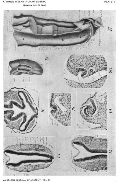
Fig. 13. A segment of the model (Fig. 1) from Secs. 5 to 20, showing: The total foldings obl. 7, 8, 9 of the neural tube dorsad of the roots of the Xth, Xlth, and Ist cervical nerves (cf. Figs. 3-4); the foldings of the skin corresponding with those of the neural tube; the metatela rapidly widening from the cephalic end of the cut surface (cf. Fig. 14); the close connection of neural and epidermic epithelium.
Fig. 14. From a photograph (X 47%) of Sec. 25 of the human embryo 148 (cf. Figs. 1, 2, 4). It shows: The neural tube just at the ventral border of the folds, obl. 8, 9, represented in Fig. 13; the cephalic, Ist, root of N. XI, attached to the base of fold, obl. 8; the 2d root of N. XI, attached to obl. 9; the Xlth N. as it passes through the 1st cervical and Froriep’s ganglia; the intimate union of the 2d and 3d cervical ganglia.
The cilia lining the tube appear faintly and stop short of the dorsal margin of the neural tube.
At the right, the first four myotomes are seen, the 4th showing especially well the dorsal division into two separate horns. Noticeable is a continuation cephalad of similar cell-groups and epidermal corrugations representing remnants of still more cephalic, occipital myotomes.
Fig. 15. A photograph (>< 473/2) of part of Sec. 44 (cf. Figs. 3, 4), showing the neural tube with its cilia, metatela, and the relations of the Xth, Xlth, and Xllth nerves.
At the left of Fig. 14, the fold, obl. 7, is seen. At the right, it has disappeared, to reappear as a more marked depression at the lower level of Fig. 15, where the attachment of the Xth N. is found.
At the left is seen the appearance of the 1st, 2d, and 3d myotomes at a lower level than in Fig. 14, and at the right, at a still lower level, as they recede farther from the skin and where the nuclei, related to the developing muscle fibers, form a band across the myotome. At the left is a continuation cephalad of the same segmented appearance of the epidermis as that which lies over the myotome.
Fig. 16. A photograph (X47175) of Sec. 192, through the neuropore to "show: The point of most intimate union of the thickened epidermis with the neural epithelium (for the extent of the neuroporic thickening, (cf. Figs. 1-8); the total folds entering into the formation of the visual lobe; the sharp angle formed by the albicantial furrow at the right; the external filling up of this angle at the left, indicating its independent ending (see text).; the mesoderm continuous over the eye-vesicle, but interrupted at the neuropore (cf. Fig. 4). The cilia. present in this section do not show clearly here.
Fig. 17. Ventral view of a large model of the nephric region of Homo 148, extending from See. 94 to Sec. 295 (X 75). It shows the two Wolflian ridges, each including the pronephric remnant; the mesonephros; the dorsal portion of the mesentery; a part of the stomach and lesser peritoneal cavity; the union of the allantoic stalk with the intestine; the cloaca and the imperforate anal plate; the right Wolffian duct and its union with the cloaca.
The opening of the pronephric tubules (Fig. 18) on the cmlomic surface is shown. Crosses indicate the position of the first eight mesonephric tubules which do not open on the caelomic surface (Fig. 19) but connect with the Wolflian duct. The 9th to the 20th tubules have no connection with the Wolflian duct. The 9th and the 13th to the 18th are open to the cualom. The 10th to the 12th are hollow but connect neither with duct nor ouelom. The 19th to the 21st are thickenings touching the ctnlomic epithelium but showing no cavity. The general arrangement of the tubules of the other side is similar but not identical.
The slight furrows at the cephalic end may represent a rudiment of the supra-renal or adrenal body.
Fig. 18. From a photograph (X 160) of Sec. 104, Homo 148, showing the pronephric tubule of the right side (Fig. 17) with its opening into the coelom and its duct traceable for a few sections farther cephalad. The cardinal vein is also seen.
Fig. 19. From a photograph (X 160) of Sec. 166 through the 7th mesonephric tubule of the right side (Fig. 17), showing the small rudiment of the glomerulus; the typical S-shaped tubule connecting with the Wolifian duct and thin-walled Bowman's capsule which a few sections farther caudad unites with the cuelomic epithelium and forms a small opening. The remainder of the first eight tubules are of this ame general type but are completely separated from the cualomic epithelium.
Fig. 20. From a photograph (X 160) of Sec. 195, through the 10th meso nephric tubule of the left side (Fig. 17), showing a wide opening to the coelom and its independence of the Wolflian duct.
The photographs reproduced on this plate were made by Henry Phelps Gage and are part of a complete series of 96 taken from the sections.
| Historic Disclaimer - information about historic embryology pages |
|---|
| Pages where the terms "Historic" (textbooks, papers, people, recommendations) appear on this site, and sections within pages where this disclaimer appears, indicate that the content and scientific understanding are specific to the time of publication. This means that while some scientific descriptions are still accurate, the terminology and interpretation of the developmental mechanisms reflect the understanding at the time of original publication and those of the preceding periods, these terms, interpretations and recommendations may not reflect our current scientific understanding. (More? Embryology History | Historic Embryology Papers) |
Reference
Gage SP. A three weeks' human embryo, with especial reference to the brain and nephric system. (1905) Amer. J Anat. 4: 409-443.
Cite this page: Hill, M.A. (2024, April 24) Embryology Gage1905-plate05.jpg. Retrieved from https://embryology.med.unsw.edu.au/embryology/index.php/File:Gage1905-plate05.jpg
- © Dr Mark Hill 2024, UNSW Embryology ISBN: 978 0 7334 2609 4 - UNSW CRICOS Provider Code No. 00098G[[[Category:Carnegie Stage 13]]
File history
Click on a date/time to view the file as it appeared at that time.
| Date/Time | Thumbnail | Dimensions | User | Comment | |
|---|---|---|---|---|---|
| current | 13:30, 18 August 2016 |  | 979 × 1,500 (271 KB) | Z8600021 (talk | contribs) | ===Plate V=== Fig. 13. A segment of the model (Fig. 1) from Secs. 5 to 20, showing: The total foldings obl. 7, 8, 9 of the neural tube dorsad of the roots of the Xth, Xlth, and Ist cervical nerves (cf. Figs. 3-4); the foldings of the skin correspondi... |
You cannot overwrite this file.
File usage
The following page uses this file:
