File:Gage1905-plate03.jpg

Original file (1,042 × 1,500 pixels, file size: 238 KB, MIME type: image/jpeg)
Plate III
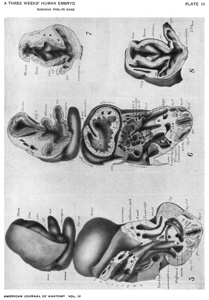
Fig. 5. A ventral view of a segment of the model (Fig. 1) extending from section 247 to 155.
The left side of the head is dissected away to show: The relation of the visual lobe, the neuropore, the olfactory and cerebral regions, and the roof of the diencephal.
The right side of the head shows: The thickened epithelium of the neuropore; the olfactory region and its extension over the cerebrum; the future lens; the mandible and gill-cleft-like pocket at the corner of the mouth.
The caudal portion of the figure (Sec. 247) shows the 23d myotome apparently continuous with the mesoderm of the leg-bud; the division of the aorta into the umbilical branches, the left one looping over the caudal end of the coelom; the left umbilical vein with a branch from the leg; the right plexiform umbilical vein passing along the body-wall toward the heart.
The middle part shows: The wide umbilicus turning to the right and containing the thick-walled vitelline sac with its veins and arteries and its union with the caudal intestine inclosed in mesentery; the reappearance of the intestine in section near the union of the Wolflian duct and allantoic stalk (cf. Fig. 17).
Fig. 6. A ventral view of a deeper segment of the model. It extends from section 185 to 155. In the head region (of. Figs. 2-5) it is cut through; the neuropore; the eye vesicles, partially constricted off from the stalk; the albicantial folds (of. Fig. 9); and the cephalic end of the thick-walled mesencephal as it dips into the albicantial region. At N. III is a strand of mesodermic tissue but apparently no true nerve fibers.
The caudal part of the figure (Sec. 185) shows: On one side, the appearance of a. myotome cut through the middle; on the other side the overlapping ends of two myotomes; the 8th mesonephric tubule opening into the Wolflian duct and with its cap of thickened peritoneal epithelium (cf. Fig. 17) and its artery and vein.
In the middle portion are: The umbilical veins at either side, the left showing the greatly divided sinuses; the vitelline veins as they approach on either side of the alimentary canal; the vitelline artery; the liver near its caudal part embedded in the transverse septum and near the level where the bile duct unites with the duodenum; the umbilicus turning to the right of the specimen.
The heart (Sec. 168) cut near its middle shows: The undivided chamber of the ventricle; a strong fold arising between the two sides and separating the exit of the bulbus arteriosus (cf. Fig. 12) from the entrance of the auriculo-ventricular canal (cf. Fig. 10).
Fig. 7. From a segment of the model cut at section 200, near the edge of the neuropore, looking into the roof of the curved brain tube and showing that the striatal, olfactory, and cerebral folds, and those of the roof of the diencephal and of the mesencephal, meet in the mid-dorsal line, there being no division by a middle partition.
Fig. 8. A segment of the model extending from Sec. 198 to 155. It cuts the neuropore (cf. Fig. 16), showing a pit on its neural aspect, and looks into the visual lobe and eye vesicles in the opposite direction from Fig. 7. It shows the notch at the tip of the optic vesicle, apparently the beginning of the optic cup.
The deep projection of the floor of the mesencephal into the albicantial region is here shown and the independence of the albicantial folds from those dorsad of it (cf. Fig. 9).
| Historic Disclaimer - information about historic embryology pages |
|---|
| Pages where the terms "Historic" (textbooks, papers, people, recommendations) appear on this site, and sections within pages where this disclaimer appears, indicate that the content and scientific understanding are specific to the time of publication. This means that while some scientific descriptions are still accurate, the terminology and interpretation of the developmental mechanisms reflect the understanding at the time of original publication and those of the preceding periods, these terms, interpretations and recommendations may not reflect our current scientific understanding. (More? Embryology History | Historic Embryology Papers) |
Reference
Gage SP. A three weeks' human embryo, with especial reference to the brain and nephric system. (1905) Amer. J Anat. 4: 409-443.
Cite this page: Hill, M.A. (2024, April 19) Embryology Gage1905-plate03.jpg. Retrieved from https://embryology.med.unsw.edu.au/embryology/index.php/File:Gage1905-plate03.jpg
- © Dr Mark Hill 2024, UNSW Embryology ISBN: 978 0 7334 2609 4 - UNSW CRICOS Provider Code No. 00098G[[[Category:Carnegie Stage 13]]
File history
Click on a date/time to view the file as it appeared at that time.
| Date/Time | Thumbnail | Dimensions | User | Comment | |
|---|---|---|---|---|---|
| current | 13:21, 18 August 2016 |  | 1,042 × 1,500 (238 KB) | Z8600021 (talk | contribs) | ===Plate III=== Fig. 5. A ventral view of a segment of the model (Fig. 1) extending from section 247 to 155. The left side of the head is dissected away to show: The relation of the visual lobe, the neuropore, the olfactory and cerebral regions, an... |
You cannot overwrite this file.
File usage
The following page uses this file:
