File:Gage1905-plate02.jpg
Original file (1,007 × 1,500 pixels, file size: 221 KB, MIME type: image/jpeg)
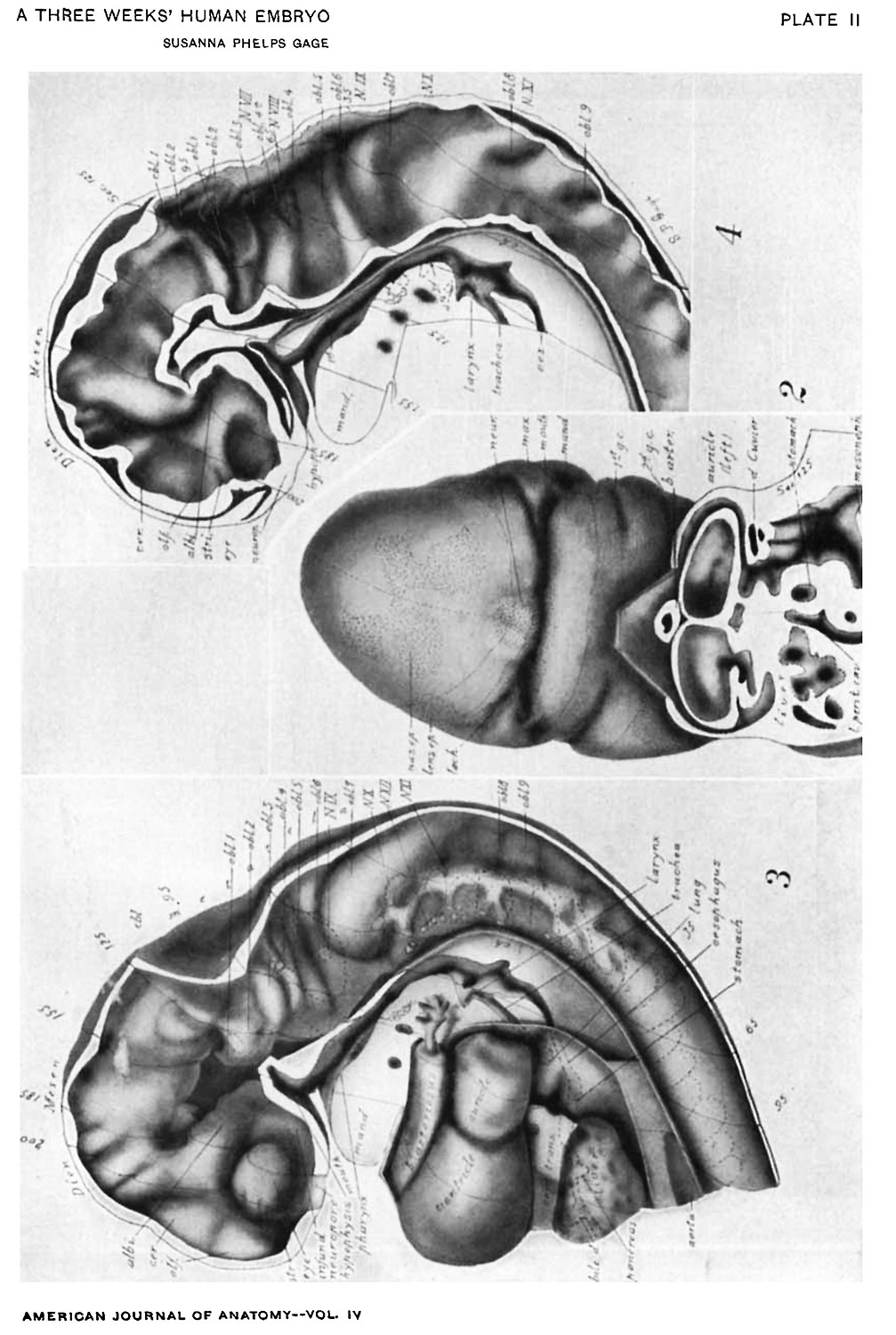
Plate II
Fig. 2. A face view of the head. As shown by the topographic lines, it is tilted to give a clear view of the parts about the mouth which is merely a wide slit between the hypophysial region of the head (cf. Figs. 3, 4) and the mandibular process.
There are seen: The small maxillary process with the depression at the corner of the mouth lying between maxilla and mandible; the H-shape of the nasal epithelium extending also over the cerebral region; the large neuroporic thickening; the lens epithelium with a tract extending along the lachrymal furrow.
The cut surface (see. 125) shows: The division in the dorsal part of the auricles (cf. Fig. 3); the entrance of the sinus venosus into the right part (left of Fig.); the liver lying in the septum transversum; the folds about the duct of Cuvier, pushing across the space to help form the diaphragm; the connection of pericardial and abdominal ccelom; the opening of the lesser peritoneal cavity into the abdominal ccelom.
Fig. 3. A view from the left side showing the central nervous system, pharynx, heart, lung, and liver. The lateral wall has been removed. Projections of the ear vesicle and myotomes 1-15 are indicated by dotted outlines.
The approximately uniform tube formed by the central nervous system and the strong cephalic flexure characteristic of this stage of development are evident. The mesoderm in the flexure has been removed.
The brain shows: The series of total folds; the great prominence of the albicantial; the relation of the neuropore to the epidermis and to the visual lobe or eye; the small size of the striatal, olfactory, and cerebral folds; the relation of the fold, oblongata 2, to the Vth nerve root (shown by a dotted circle); of the fold, obl. 4, to the VIIth and VIIIth nerves (dotted circles); of fold, obl. 6 to N. IX; the great size of fold, obl. 7 and its relation to N. X; the continuity and segmented character of the ganglionic chain in the neck region with the roots of the XIth nerve extending along its dorsal side; and the folds in the myel.
There are seen: The inner tube of the bulbus arteriosus as it enters the floor of the pharynx and divides into the aortic arches; the median thyroid cephalad of this branching; lateral folds just cephalad of the thyroid, the only rudiments of the tongue present; the bursa pharyngea, the dorsal pocket at the division of trachea and esophagus (at left of abbreviation ch.); dotted lines indicating the outline of the epithelial tubes forming lung and alimentary canal; the wide communication of the pericardial and abdominal ctelom dorsad of the septum transversum; the point of union of the aortae (aorta); the hypophysis cut to the left of the middle line.
Fig. 4. A mesal view of the brain, myel, and pharynx. The brain shows from the interior the same total folds as Fig. 3, but brings out somewhat more clearly the grouping of folds into lobules (cf. Table II, in the text). The elevations in Fig. 3 correspond to the depressions in Fig. 4.
Especially noticeable in this figure are: The short striatal folds; the apparent continuity of the albicantial fold with the roof fold (cf. Figs. 8, 9); the cerebellar folds 1 and 2 (cf. Fig. 11, cbl.); the cleft in the oblongata fold 4, on either side of which arise the roots of the Vllth and VIIIth nerves (shown by dotted circles); the cut ends of the ring of mesoderm surrounding the neuropore; the intrusion of mesoderm into the cephalic flexure; the fact that no mesoderm intervenes between hypophysis and infundibulum on the middle line; the union of all layers in the roof of the oblongata; the dark areas in the pharynx, indicating with their dotted extension the membranous parts of the 1st to the 4th gill pouches; the notochord touching the caudal wall of the hypophysis and coming in close contact with the roof of the pharynx between the level of the 1st and 3d gill pouches.
| Historic Disclaimer - information about historic embryology pages |
|---|
 |
Reference
Gage SP. A three weeks' human embryo, with especial reference to the brain and nephric system. (1905) Amer. J Anat. 4: 409-443.
Cite this page: Hill, M.A. (2024, April 20) Embryology Gage1905-plate02.jpg. Retrieved from https://embryology.med.unsw.edu.au/embryology/index.php/File:Gage1905-plate02.jpg
- © Dr Mark Hill 2024, UNSW Embryology ISBN: 978 0 7334 2609 4 - UNSW CRICOS Provider Code No. 00098G[[[Category:Carnegie Stage 13]]
File history
Click on a date/time to view the file as it appeared at that time.
| Date/Time | Thumbnail | Dimensions | User | Comment | |
|---|---|---|---|---|---|
| current | 13:14, 18 August 2016 | | 1,007 × 1,500 (221 KB) | Z8600021 (talk | contribs) | ==Plate II== Fig. 2. A face view of the head. As shown by the topographic lines, it is tilted to give a clear view of the parts about the mouth which is merely a wide slit between the hypophysial region of the head (cf. Figs. 3, 4) and the mandibular... |
You cannot overwrite this file.
File usage
The following page uses this file: