File:Frazer1935 fig08.jpg

Original file (1,264 × 667 pixels, file size: 95 KB, MIME type: image/jpeg)
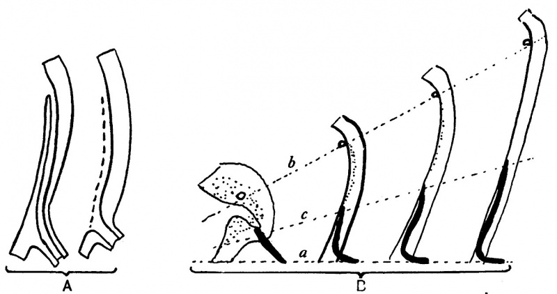
Fig. 8. These diagrams represent only the lining cell layers
A. To show the method by which the actual opening of the duct is gained. The superficial wall of the embedded part breaks down and disappears (interrupted line in second figure), leaving orifice of duct at its low level.
B. Diagrams to show the efiect on duct (black) and ureteric level of growth of sinus upwards from the fixed level (a). The ureteric orifice is raised most (b), while the embedded end of the duct is not only turned up but also lengthened (c) to a smaller extent. The epithelial mass (dotted) round openings of ureter and duct in the first figure is hollowed to a small funnel and then drawn out between b and c.
| Historic Disclaimer - information about historic embryology pages |
|---|
 |
- Links: Fig 1 Wolflian body embryo 4.9 mm | Fig 2 Walls of cavities from embryos 4.9, 5, and 8 mm | Fig 3 Cloacal expansion outwards along Wolflian duct | Fig 4 Lateral projection of sinus | Fig 5 Wolffian duct “taken up” into cavity of bladder | Fig 6 Lateral dilatation of sinus embryo 8 mm | Fig 7 Dorsal walls of urogenital sinuses embryos 18, 21, and 28 mm | Fig 8 Lining cell layers
Reference
Frazer JE. The terminal part of the wolffian duct. (1935) J Anat., 69(4): 455–468. PMID 17104551
Cite this page: Hill, M.A. (2024, April 18) Embryology Frazer1935 fig08.jpg. Retrieved from https://embryology.med.unsw.edu.au/embryology/index.php/File:Frazer1935_fig08.jpg
- © Dr Mark Hill 2024, UNSW Embryology ISBN: 978 0 7334 2609 4 - UNSW CRICOS Provider Code No. 00098G
File history
Click on a date/time to view the file as it appeared at that time.
| Date/Time | Thumbnail | Dimensions | User | Comment | |
|---|---|---|---|---|---|
| current | 15:46, 30 August 2015 | | 1,264 × 667 (95 KB) | Z8600021 (talk | contribs) | |
| 15:25, 30 August 2015 |  | 1,438 × 1,018 (249 KB) | Z8600021 (talk | contribs) |
You cannot overwrite this file.
File usage
The following page uses this file: