File:Frazer1914 fig04.jpg

Original file (1,000 × 709 pixels, file size: 129 KB, MIME type: image/jpeg)
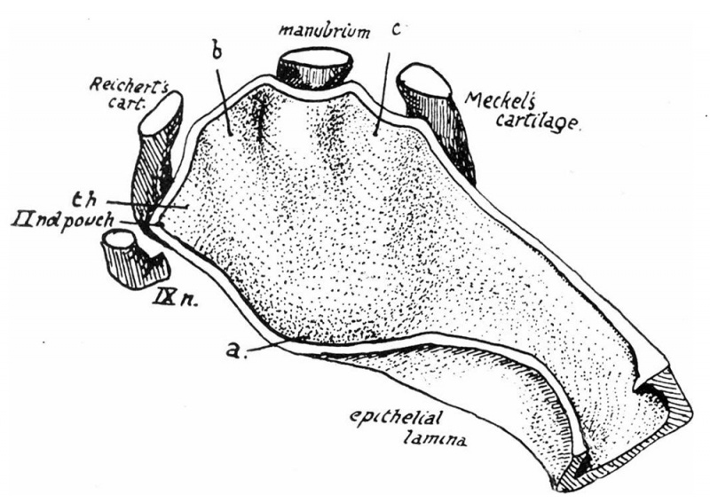
Fig. 4. Left recess simplified from model 35 mm Embryo
a, bottom of tympanum practically corresponding with lower extremity mental plate and tympanic ring;
b, c, prominences made by back and front edges of mental plate growing in round the manubrium ; th., tympano-hyal area.
Fig. 4 is from a model of a 35-mm. embryo, and shows how greatly the third arch has extended forward; comparison with the earlier stages makes this evident. The recess as a whole is slightly longer, and the growth of the third arch particularly affects the inner part and narrows it, thus definitely indicating the distinction between tube and tympanum. But it is equally plain that this narrowing has not been obtained by a raising up of the epithelial floor as a result of growth under it, but by a fusion of the surfaces of the first and third arches where they have come into contact; for an epithelial lamina is present below the newly made “ floor" in this region which is continuous with the epithelial lining of the cavity along its length from the hinder boundary of the pharyngeal opening to the hollow situated at the foot of the anterior district of the second arch in the tympanic region. Such a lamina can be nothing but the covering of the prominent arches caught between them as they meet. The lamina, as shown in the model, consists only of a solid and continuous epithelial septum; but under the microscope broken layers and masses of cells can be found which are not suitable for modelling, but indicate that the fusion. is more extensive behind and below than is suggested by the model. What is probably the beginning of this process is found in the 29—mm. embryo, in which an epithelial mass can be seen lying above the front part of the forward extension of the third arch, between the thicker posterior part of this extension and the first arch eminence.
| Historic Disclaimer - information about historic embryology pages |
|---|
 |
- Links: Fig 1 | Fig 2 | Fig 3 | Fig 4 | Fig 5 | Fig 6 | 1914 Frazer | Pharyngeal arches | Historic Embryology Papers
Reference
Frazer JE. The second visceral arch and groove in the tubo-tympanic region. (1914) J Anat Physiol. 48(4): 391-408. PMID 17233005
Cite this page: Hill, M.A. (2024, April 25) Embryology Frazer1914 fig04.jpg. Retrieved from https://embryology.med.unsw.edu.au/embryology/index.php/File:Frazer1914_fig04.jpg
- © Dr Mark Hill 2024, UNSW Embryology ISBN: 978 0 7334 2609 4 - UNSW CRICOS Provider Code No. 00098G
File history
Click on a date/time to view the file as it appeared at that time.
| Date/Time | Thumbnail | Dimensions | User | Comment | |
|---|---|---|---|---|---|
| current | 06:12, 9 January 2017 | | 1,000 × 709 (129 KB) | Z8600021 (talk | contribs) | |
| 06:12, 9 January 2017 | 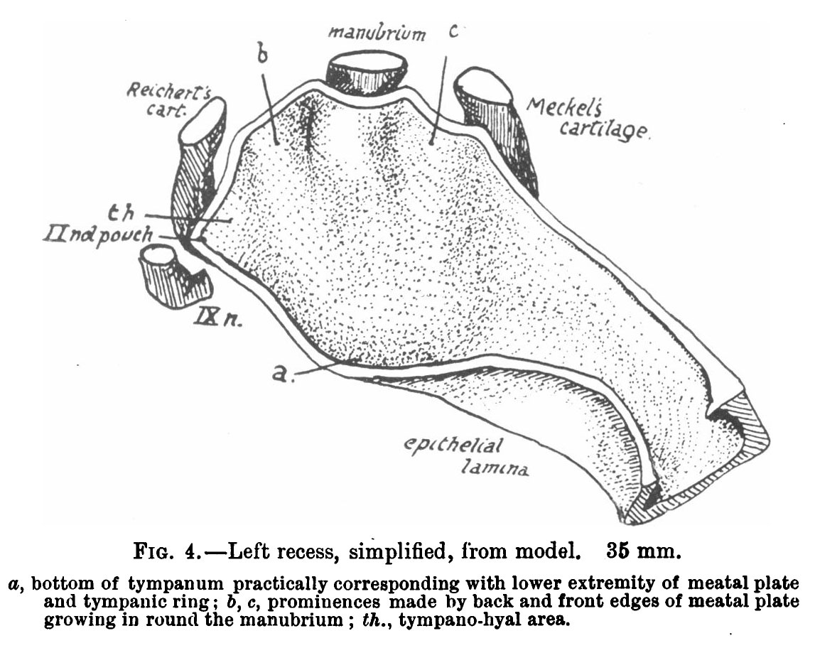 | 1,147 × 912 (174 KB) | Z8600021 (talk | contribs) | {{Historic Disclaimer}} ===Reference=== {{Ref-Frazer1914}} {{Footer}} |
You cannot overwrite this file.
File usage
The following page uses this file: