File:Frazer1910 fig11.jpg

Original file (755 × 702 pixels, file size: 70 KB, MIME type: image/jpeg)
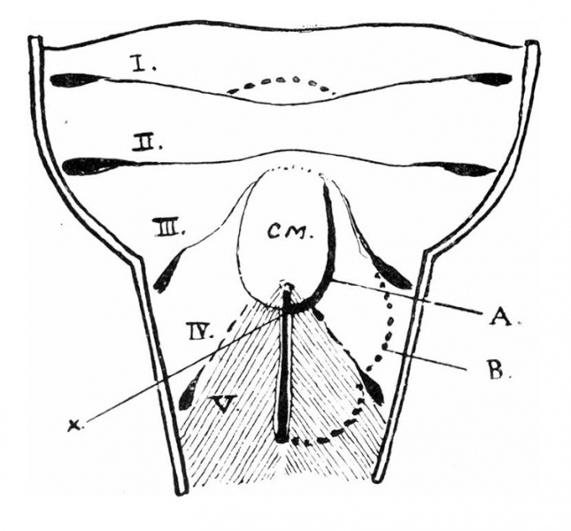
Fig. 11. The diagram shows the increased obliquity of the hinder arches
The pulmonary opening is thus apparently in front of its position in the lust figure. The interrupted line B shows the outline of the lateral mass, which thus comprises 4th and 5th arches, and the thick line A indicates the line at which it begins to overlap the central mass; this line is seen to correspond with the margin of the central mass.
The front end of the pulmonary opening is on the base of the central mass, bordered by the “ original apices ” of the 5th arches, and the line of overlapping therefore crosses the sagittal cleft at the point X; therefore, when overlapping occurs, the sagittal line in front of X is left on the base of the central mass with the original apex on each side of it, and a new dorsal apex of the 5th arch is lifted over the central mass, being originally behind and outside X.
The 4th arch element of the lateral mass is seen spreading up the side of C. M. and being lost there, and the 3rd arch is clear across the middle line.
The continuity between the two “ apices” of each 5th nrch mass must lie under the sulcus formed at the line A where the shading crosses it.
| Historic Disclaimer - information about historic embryology pages |
|---|
 |
- Links: fig 1 | fig 2 | fig 3 | fig 4 | fig 5 | fig 6 | fig 7 | fig 8 | fig 9 | fig 10 | fig 11 | fig 12 | fig 13 | fig 14 | fig 15 | fig 16 | fig 17 | fig 18 | fig 19 | 1910 Frazer | Historic Embryology Papers | Respiratory System Development
Reference
Frazer JE. Development of the larynx. (1910) J Anat. 44: 156-191. PMID 17232839
Cite this page: Hill, M.A. (2024, April 16) Embryology Frazer1910 fig11.jpg. Retrieved from https://embryology.med.unsw.edu.au/embryology/index.php/File:Frazer1910_fig11.jpg
- © Dr Mark Hill 2024, UNSW Embryology ISBN: 978 0 7334 2609 4 - UNSW CRICOS Provider Code No. 00098G
File history
Click on a date/time to view the file as it appeared at that time.
| Date/Time | Thumbnail | Dimensions | User | Comment | |
|---|---|---|---|---|---|
| current | 09:35, 11 January 2017 | | 755 × 702 (70 KB) | Z8600021 (talk | contribs) | |
| 09:34, 11 January 2017 | 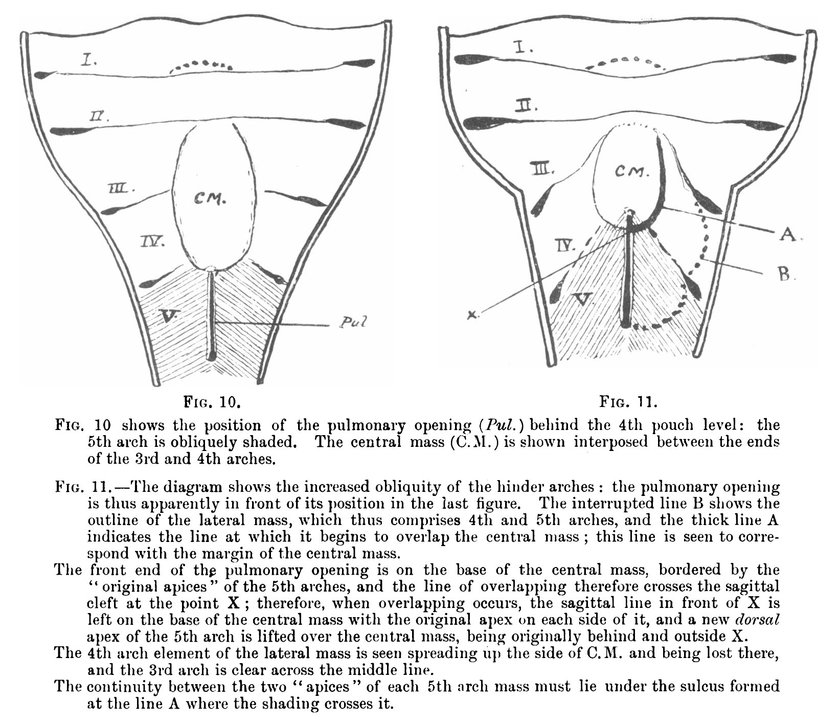 | 1,643 × 1,416 (400 KB) | Z8600021 (talk | contribs) | {{Frazer1910 figures}} |
You cannot overwrite this file.
File usage
The following 2 pages use this file: