File:Foster118b.jpg
Original file (809 × 1,077 pixels, file size: 232 KB, MIME type: image/jpeg)
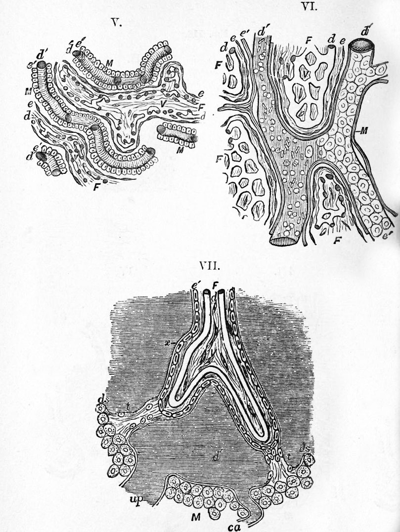
Fig. 118. Histology of the placenta. Diagrammatic representations of the minute structure of the placenta
From Turner. See also 118a
VI. Structure of placenta of a Sloth.
- On the right side of the figure the flat maternal epithelial cells are shewn in situ. On the left side they are removed, and the dilated maternal vessel with its blood-corpuscles is exposed.
VII. Structure of Human placenta.
- In addition to the letters already referred to, ds, ds. represents the decidua serotina of the placenta ; t, t. trabeculae of serotina passing to the foetal villi ; ca. curling artery ; up. utero-placental vein ; x. a prolongation of maternal tissue on the exterior of the villus outside the cellular layer e', which may represent either the endothelium of the maternal blood-vessel or delicate connective tissue belonging to the serotina, or both. The layer e' represents maternal cells derived from the serotina. The layer of foetal epithelium cannot be seen on the villi of the fully-formed human placenta.
In the human placenta (VII.), as in that of Apes, the greatest modification is found. Here the maternal vessels have completely lost their capillary form, and have become expanded into large freely communicating sinuses (d f ). In these sinuses the foetal villi hang for the most part freely, though occasionally attached to their walls by strands of tissue (t). In the late stages of fcetal life there is only one epithelial layer (e} between the maternal and fcetal vessels, which closely invests the fcetal villi, but is part of the uterine tissue. In the foetal villi the vessels retain their capillary form.
Legend
F. the foetal ; M. the maternal placenta ; e. epithelium of chorion ; e'. epithelium of maternal placenta ; d. foetal bloodvessels ; d'. maternal blood-vessels ; v. villus.
Reference
Foster, M., Balfour, F. M., Sedgwick, A., & Heape, W. (1883). The Elements of Embryology. (2nd ed.). London: Macmillan and Co.
- Volume 1 - The History of the Chick: Egg structure and incubation beginning | Summary whole incubation | First day | Second day - first half | Second day - second half | Third day | Fourth day | Fifth day | Sixth day to incubation end | Figures 1
- Volume 2 - The History of the Mammalian Embryo: General Development | Embryonic Membranes and Yolk-Sac | Organs from Epiblast | Organs from Mesoblast | Alimentary Canal | Appendix | Figures 2
| Historic Disclaimer - information about historic embryology pages |
|---|
 |
File history
Click on a date/time to view the file as it appeared at that time.
| Date/Time | Thumbnail | Dimensions | User | Comment | |
|---|---|---|---|---|---|
| current | 18:12, 12 January 2011 | | 809 × 1,077 (232 KB) | S8600021 (talk | contribs) | {{Template:Foster_1883_Figures_2}} Category:Cartoon |
You cannot overwrite this file.
File usage
The following 2 pages use this file: