File:Foster115.jpg
From Embryology
Size of this preview: 606 × 600 pixels. Other resolution: 847 × 838 pixels.
Original file (847 × 838 pixels, file size: 131 KB, MIME type: image/jpeg)
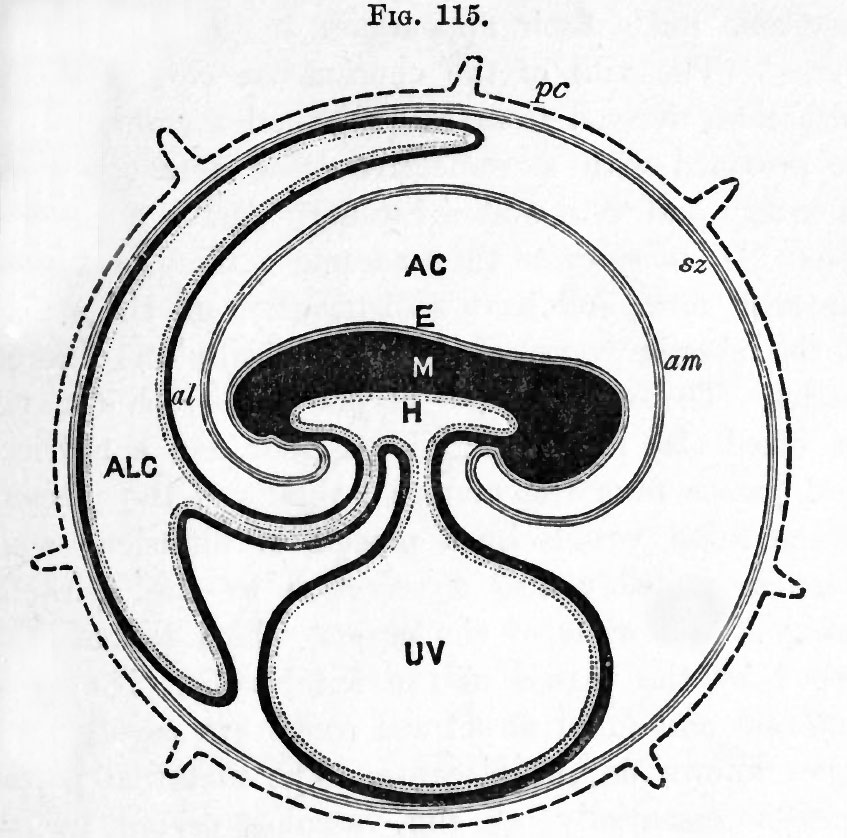
Fig. 115. Diagram of the foetal membranes of a mammal
From Turner.
Structures which either are or have been at an earlier period of development continuous with each other are represented by the same character of shading.
Legend
pc. zona with villi ; sz. subzonal membrane ; E. epiblast of embryo ; am. amnion ; AC. amniotic cavity ; M. mesoblast of embryo ; H. hypoblast of embryo ; UV. umbilical vesicle ; al. allantois ; ALC. allantoic cavity.
Reference
Foster, M., Balfour, F. M., Sedgwick, A., & Heape, W. (1883). The Elements of Embryology. (2nd ed.). London: Macmillan and Co.
- Volume 1 - The History of the Chick: Egg structure and incubation beginning | Summary whole incubation | First day | Second day - first half | Second day - second half | Third day | Fourth day | Fifth day | Sixth day to incubation end | Figures 1
- Volume 2 - The History of the Mammalian Embryo: General Development | Embryonic Membranes and Yolk-Sac | Organs from Epiblast | Organs from Mesoblast | Alimentary Canal | Appendix | Figures 2
| Historic Disclaimer - information about historic embryology pages |
|---|
 |
File history
Click on a date/time to view the file as it appeared at that time.
| Date/Time | Thumbnail | Dimensions | User | Comment | |
|---|---|---|---|---|---|
| current | 18:11, 12 January 2011 | | 847 × 838 (131 KB) | S8600021 (talk | contribs) | FIG. 115. DIAGRAM OF THE FCETAL MEMBRANES OP A MAMMAL. (From Turner.) Structures which either are or have been at an earlier period of development continuous with each other are represented by the same character of shading. pc. zona with villi ; sz. subz |
You cannot overwrite this file.
File usage
The following 2 pages use this file: