File:Foster102.jpg
Original file (945 × 458 pixels, file size: 90 KB, MIME type: image/jpeg)
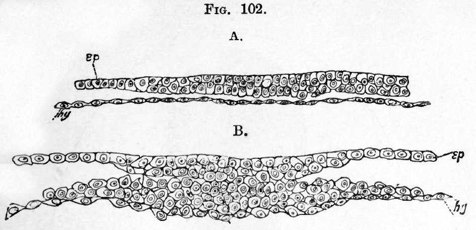
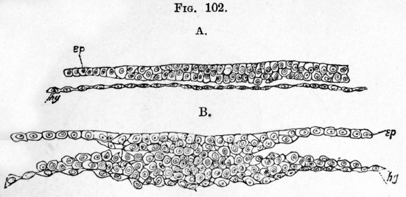
FIG. 102. Two TRANSVERSE SUCTIONS THROUGH THE EMBRYONIC AREA OF AN EMBRYO RABBIT OF SEVEN DAYS.
The embryo has nearly the appearance represented in Fig. 100.
A. is taken through the anterior part of the embryonic area. It represents about half the breadth of the area, and there is no trace of a medullary groove or of the mesoblast.
B. is taken through the posterior part of the primitive streak. ep. epiblast ; hy. hypoblast.
Reference
Foster, M., Balfour, F. M., Sedgwick, A., & Heape, W. (1883). The Elements of Embryology. (2nd ed.). London: Macmillan and Co.
- Volume 1 - The History of the Chick: Egg structure and incubation beginning | Summary whole incubation | First day | Second day - first half | Second day - second half | Third day | Fourth day | Fifth day | Sixth day to incubation end | Figures 1
- Volume 2 - The History of the Mammalian Embryo: General Development | Embryonic Membranes and Yolk-Sac | Organs from Epiblast | Organs from Mesoblast | Alimentary Canal | Appendix | Figures 2
| Historic Disclaimer - information about historic embryology pages |
|---|
 |
File history
Click on a date/time to view the file as it appeared at that time.
| Date/Time | Thumbnail | Dimensions | User | Comment | |
|---|---|---|---|---|---|
| current | 18:04, 12 January 2011 | | 945 × 458 (90 KB) | S8600021 (talk | contribs) | FIG. 102. Two TRANSVERSE SUCTIONS THROUGH THE EMBRYONIC AREA OF AN EMBRYO RABBIT OF SEVEN DAYS. The embryo has nearly the appearance represented in Fig. 100. A. is taken through the anterior part of the embryonic area. It represents about half the bread |
You cannot overwrite this file.
File usage
The following 2 pages use this file: