File:Foster050.jpg
Foster050.jpg (792 × 327 pixels, file size: 43 KB, MIME type: image/jpeg)
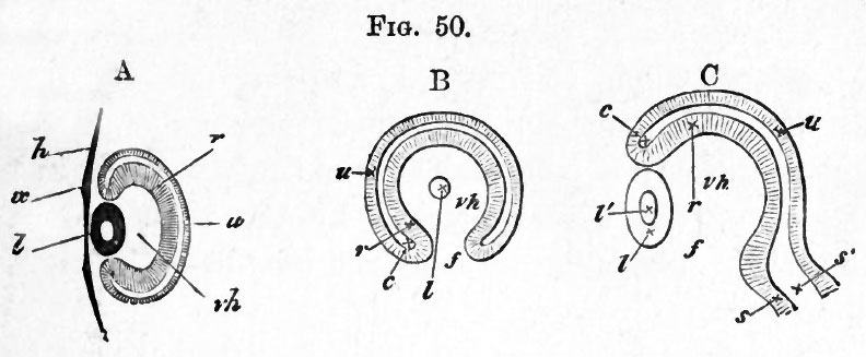
FIG. 50.
A. Diagrammatic section taken perpendicular to the plane of the paper, along the line y, y, Fig. 49. The stalk is not seen, the* section falling quite out of its region, vh, hollow of optic cup filled with vitreous humour ; other letters as in Fig. 47 B.
B. Section taken parallel to the plane of paper through Fig. 49, so far behind the front surface of the eye as to shave off a small portion of the posterior surface of the lens I, but so far in front as not to be carried at all through the stalk. Letters as before ; /, the choroidal fissure.
C. Section along the line z, z, perpendicular to the plane of the paper, to shew the choroidal fissure /, and the continuity of the cavity of the optic stalk with that of the primary optic vesicle. Had this section been taken a little to either side of the line z, 2, the wall of the optic cup would have extended up to the lens below as well as above. Letters as above.
| Historic Disclaimer - information about historic embryology pages |
|---|
 |
Reference
Foster, M., Balfour, F. M., Sedgwick, A., & Heape, W. (1883). The Elements of Embryology. (2nd ed.). London: Macmillan and Co.
The Elements of Embryology (1883)
File history
Click on a date/time to view the file as it appeared at that time.
| Date/Time | Thumbnail | Dimensions | User | Comment | |
|---|---|---|---|---|---|
| current | 10:17, 11 January 2011 | | 792 × 327 (43 KB) | S8600021 (talk | contribs) | FIG. 50. A. Diagrammatic section taken perpendicular to the plane of the paper, along the line y, y, Fig. 49. The stalk is not seen, the* section falling quite out of its region, vh, hollow of optic cup filled with vitreous humour ; other letters as in F |
You cannot overwrite this file.
File usage
The following 2 pages use this file: