File:Foster044.jpg
Foster044.jpg (461 × 560 pixels, file size: 55 KB, MIME type: image/jpeg)
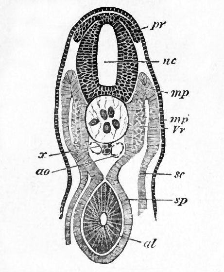
FIG. 44. TRANSVERSE SECTION THROUGH THE TRUNK OF A YOUNG EMBRYO OF A DOG-FlSH.
nc. neural canal ; pr. posterior root of spinal nerve ; x. subnotochordal rod ; ao. aorta ; sc. somatic mesoblast ; sp. splanchnic mesoblast ; mp. muscle-plate ; mp'. portion of muscle-plate converted into muscle ; Vv. portion of the vertebral plate which will give rise to the vertebral bodies ; al. alimentary tract.
| Historic Disclaimer - information about historic embryology pages |
|---|
 |
Reference
Foster, M., Balfour, F. M., Sedgwick, A., & Heape, W. (1883). The Elements of Embryology. (2nd ed.). London: Macmillan and Co.
The Elements of Embryology (1883)
File history
Click on a date/time to view the file as it appeared at that time.
| Date/Time | Thumbnail | Dimensions | User | Comment | |
|---|---|---|---|---|---|
| current | 09:49, 11 January 2011 | | 461 × 560 (55 KB) | S8600021 (talk | contribs) | FIG. 44. TRANSVERSE SECTION THROUGH THE TRUNK OF A YOUNG EMBRYO OF A DOG-FlSH. nc. neural canal ; pr. posterior root of spinal nerve ; x. subnotochordal rod ; ao. aorta ; sc. somatic mesoblast ; sp. splanchnic mesoblast ; mp. muscle-plate ; mp'. portion |
You cannot overwrite this file.
File usage
The following 2 pages use this file: