File:Foster034.jpg

Original file (1,003 × 717 pixels, file size: 153 KB, MIME type: image/jpeg)
FIG. 34. TRANSVERSE SECTION THROUGH THE DORSAL REGION OF AN EMBRYO OF 45 HOURS.
A. epiblast. B. mesoblast. C. hypoblast consisting of a single row of flattened cells. M. c. medullary canal. P. v. mesoblastic somite. W. d. Wolffian duct. S. o. Somatopleure. S.p. Splanchnopleure. p.p. pleuroperitoneal cavity, c. h. notochord. a. o. dorsal aorta, v. blood-vessels of the yolksac, o.p. line of junction between opaque and pellucid areas ; w. palisade-like yolk spheres which constitute the germinal wall.
Only one-half of the section is represented in the figure if completed it would be bilaterally symmetrical about the line of the medullary canal.
| Historic Disclaimer - information about historic embryology pages |
|---|
 |
Reference
Foster, M., Balfour, F. M., Sedgwick, A., & Heape, W. (1883). The Elements of Embryology. (2nd ed.). London: Macmillan and Co.
The Elements of Embryology (1883)
File history
Click on a date/time to view the file as it appeared at that time.
| Date/Time | Thumbnail | Dimensions | User | Comment | |
|---|---|---|---|---|---|
| current | 08:47, 11 January 2011 | | 1,003 × 717 (153 KB) | S8600021 (talk | contribs) | |
| 08:45, 11 January 2011 | 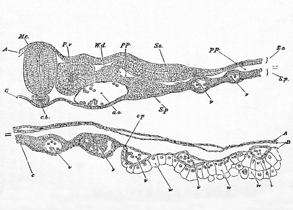 | 1,003 × 717 (153 KB) | S8600021 (talk | contribs) | {{Template:Foster 1883 Figures}} |
You cannot overwrite this file.
File usage
The following 2 pages use this file: