File:Foster032.jpg
From Embryology
No higher resolution available.
Foster032.jpg (542 × 395 pixels, file size: 48 KB, MIME type: image/jpeg)
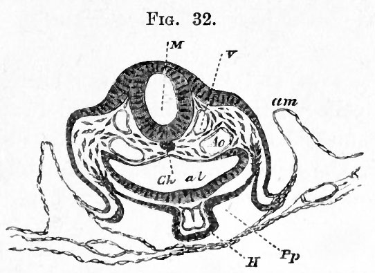
FIG. 32. TRANSVERSE SECTION OF AN EMBRYO AT THE END OF THE SECOND DAY PASSING THROUGH THE REGION OF THE BULBUS ARTERIOSUS. (Copied from His.)
M. medullary canal in the region of the hind brain ; V. anterior cardinal vein ; Ao. Aorta ; Ch. Notochord ; al. alimentary canal ; H. Heart (bulbus arteriosus) ; Pp. Pleuroperitoneal cavity; am. amnion.
| Historic Disclaimer - information about historic embryology pages |
|---|
 |
Reference
Foster, M., Balfour, F. M., Sedgwick, A., & Heape, W. (1883). The Elements of Embryology. (2nd ed.). London: Macmillan and Co.
The Elements of Embryology (1883)
File history
Click on a date/time to view the file as it appeared at that time.
| Date/Time | Thumbnail | Dimensions | User | Comment | |
|---|---|---|---|---|---|
| current | 08:04, 9 January 2011 | | 542 × 395 (48 KB) | S8600021 (talk | contribs) | FIG. 32. TRANSVERSE SECTION OF AN EMBRYO AT THE END OF THE SECOND DAY PASSING THROUGH THE REGION OF THE BULBUS ARTERIOSUS. (Copied from His.) M. medullary canal in the region of the hind brain ; V. anterior cardinal vein ; Ao. Aorta ; Ch. Notochord ; al. |
You cannot overwrite this file.
File usage
The following 3 pages use this file: