File:Foster017.jpg
From Embryology

Size of this preview: 800 × 339 pixels. Other resolution: 947 × 401 pixels.
Original file (947 × 401 pixels, file size: 72 KB, MIME type: image/jpeg)
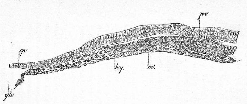
FIG. 17. TRANSVERSE SECTION THROUGH THE FRONT END OP THE PRIMITIVE STREAK OF A BLASTODERM OF THE SAME AGE AS FIG. 16.
pv. primitive groove ; m. mesoblast ; ep. epiblast ; hy. hypoblast ; yh. yolk of germinal wall.
| Historic Disclaimer - information about historic embryology pages |
|---|
 |
Reference
Foster, M., Balfour, F. M., Sedgwick, A., & Heape, W. (1883). The Elements of Embryology. (2nd ed.). London: Macmillan and Co.
The Elements of Embryology (1883)
File history
Click on a date/time to view the file as it appeared at that time.
| Date/Time | Thumbnail | Dimensions | User | Comment | |
|---|---|---|---|---|---|
| current | 07:01, 9 January 2011 | | 947 × 401 (72 KB) | S8600021 (talk | contribs) | FIG. 17. TRANSVERSE SECTION THROUGH THE FRONT END OP THE PRIMITIVE STREAK OF A BLASTODERM OF THE SAME AGE AS FIG. 16. pv. primitive groove ; m. mesoblast ; ep. epiblast ; hy. hypoblast ; yh. yolk of germinal wall. {{Template:Foster 1883 Figures}} |
You cannot overwrite this file.
File usage
The following 2 pages use this file: