File:Foster009ad.jpg

Original file (977 × 1,038 pixels, file size: 100 KB, MIME type: image/jpeg)
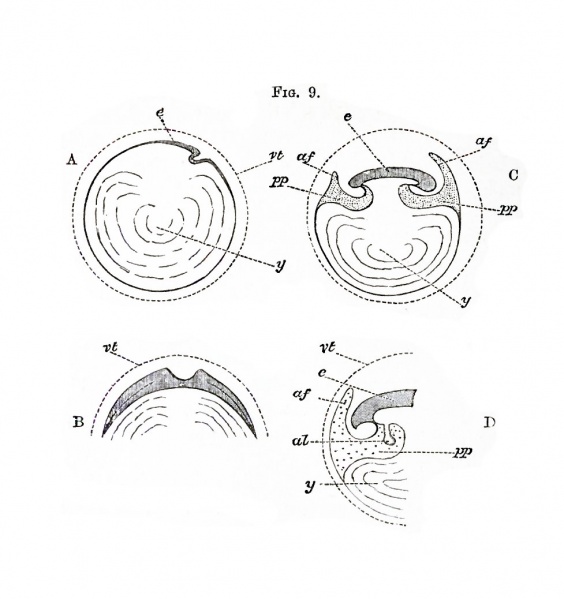
Fig. 9, A to N forms a series of purely diagrammatic representations introduced to facilitate the comprehension of the manner in which the body of the embryo is formed, and of the various relations of the yolk-sac, amnion and allantois.
A, which maybe considered as a vertical section taken longitudinally along the axis of the embryo, represents the relations of the parts of the egg at the time of the first appearance of the head-fold, seen on the right-hand side of the blastoderm e. The blastoderm is spreading both behind (to the left hand in the figure), and in front (to right hand) of the head-fold, its limits being indicated by the shading and thickening for a certain distance of the margin of the yolk y. As yet there is no fold on the left side of e corresponding to the head-fold on the right.
B is a vertical transverse section of the same period drawn for convenience sake on a larger scale (it should have been made flatter and less curved). It shews that the blastoderm (vertically shaded) is extending laterally as well as fore and aft, in fact in all directions ; but there are no lateral folds, and therefore no lateral limits to the body of the embryo as distinguished from the blastoderm. Incidentally it shews the formation of the medullary groove by the rising up of the laminae dorsales. Beneath the section of the groove is seen the rudiment of the notochord. On either side a line indicates the cleavage of the mesoblast just commencing.
In C, which represents a vertical longitudinal section of later date, both head-fold (on the right) and tail-fold (on the left) have advanced considerably. The alimentary canal is therefore closed in, both in front and behind, but is in the middle still widely open to the yolk y below. Though the axial parts of the embryo have become thickened by growth, the body- walls are still thin ; in them however is seen the cleavage of the mesoblast, and the divergence of the somatopleure and splanchnopleure. The splanchnopleure both at the head and at the tail is folded in to a greater extent than the somatopleure, and forms the still wide splanchnic stalk. At the end of the stalk, which is as yet short, it bends outwards again and spreads over the surface of the yolk. The somatopleure, folded in less than the splanchnopleure to form the wider somatic stalk, sooner bends round and runs outwards again. At a little distance from both the head and the tail it is raised up into a fold, a/, a/, that in front of the head being the highest. These are the amniotic folds. Descending from either fold, it speedily joins the splanchnopleure again, and the two, once more united into an uncleft membrane, extend some way downwards over the yolk, the limit or outer margin of the opaque area not being shewn. All the space between the somatopleure and the splanchnopleure, pp } is shaded with dots. Close to the body this space may be called the pleuroperitoneal cavity ; but outside the body it runs up into either amniotic fold, and also extends some little way over the yolk.
D represents the tail end at about the same stage on a more enlarged scale, in order to illustrate the position of the allantois al (which was for the sake of simplicity omitted in (7), shewn as a bud from the splanchnopleure, stretching downwards into the pleuroperitoneal cavity^. The dotted area representing as before the whole space between the splanchnopleure and the somatopleure, it is evident that a way is open for the allantois to extend from its present position into the space between the two limbs of the amniotic fold af.
| Historic Disclaimer - information about historic embryology pages |
|---|
| Pages where the terms "Historic" (textbooks, papers, people, recommendations) appear on this site, and sections within pages where this disclaimer appears, indicate that the content and scientific understanding are specific to the time of publication. This means that while some scientific descriptions are still accurate, the terminology and interpretation of the developmental mechanisms reflect the understanding at the time of original publication and those of the preceding periods, these terms, interpretations and recommendations may not reflect our current scientific understanding. (More? Embryology History | Historic Embryology Papers) |
Reference
Foster, M., Balfour, F. M., Sedgwick, A., & Heape, W. (1883). The Elements of Embryology. (2nd ed.). London: Macmillan and Co.
The Elements of Embryology (1883)
File history
Click on a date/time to view the file as it appeared at that time.
| Date/Time | Thumbnail | Dimensions | User | Comment | |
|---|---|---|---|---|---|
| current | 15:48, 8 January 2011 |  | 977 × 1,038 (100 KB) | S8600021 (talk | contribs) | Fig. 9, A to N forms a series of purely diagrammatic representations introduced to facilitate the comprehension of the manner in which the body of the embryo is formed, and of the various relations of the yolk-sac, amnion and allantois. A, which maybe co |
You cannot overwrite this file.
File usage
The following 2 pages use this file:
