File:Foster008.jpg

Original file (959 × 456 pixels, file size: 100 KB, MIME type: image/jpeg)
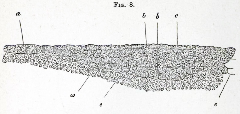
FIG. 8. SECTION OF THE GERMINAL DISC OF A FOWL DURING THE LATER STAGES OF SEGMENTATION.
The section, which represents rather more than half the breadth of the blastoderm (the middle line being shewn at c), shews that the upper and central parts of the disc segment faster than those below and towards the periphery. At the periphery the segments are still very large. One of the larger segments is shewn at a. In the majority of segments a nucleus can be seen ; and it seems probable that a nucleus is present in all. Most of the segments are filled with highly refracting spherules, but these are more numerous in some cells (especially the larger cells near the yolk) than in others. In the central part of the blastoderm the upper cells have commenced to form a distinct layer.
a. large peripheral cell.
b. larger cells of the lower parts of the blastoderm,
c. middle line of blastoderm,
e. edge of the blastoderm adjoining the white yolk.
w. white yolk.
| Historic Disclaimer - information about historic embryology pages |
|---|
 |
Reference
Foster, M., Balfour, F. M., Sedgwick, A., & Heape, W. (1883). The Elements of Embryology. (2nd ed.). London: Macmillan and Co.
The Elements of Embryology (1883)
File history
Click on a date/time to view the file as it appeared at that time.
| Date/Time | Thumbnail | Dimensions | User | Comment | |
|---|---|---|---|---|---|
| current | 15:44, 8 January 2011 | | 959 × 456 (100 KB) | S8600021 (talk | contribs) | {{Template:Foster 1883 Figures}} |
You cannot overwrite this file.
File usage
The following 2 pages use this file: