File:Foster003.jpg

Original file (1,077 × 261 pixels, file size: 39 KB, MIME type: image/jpeg)
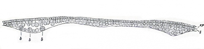
Fig. 3. Section of a blastoderm of a fowl's egg at the commencement of incubation
The thin but complete upper layer ep composed of columnar cells rests on the incomplete lower layer I, composed of larger and more granular cells.
The lower layer is thicker in some places than in others, and is especially thick at the periphery.
The line below the under layer marks the upper surface of the white yolk. The larger so-called formative cells are seen at 6, lying on the white yolk. The figure does not take in quite the whole breadth of the blastoderm ; but the reader must understand that both to the right hand and the left ep is continued farther than I, so that at the extreme edge it rests directly on the white yolk.
| Historic Disclaimer - information about historic embryology pages |
|---|
 |
Reference
Foster, M., Balfour, F. M., Sedgwick, A., & Heape, W. (1883). The Elements of Embryology. (2nd ed.). London: Macmillan and Co.
The Elements of Embryology (1883)
File history
Click on a date/time to view the file as it appeared at that time.
| Date/Time | Thumbnail | Dimensions | User | Comment | |
|---|---|---|---|---|---|
| current | 14:51, 8 January 2011 | 1,077 × 261 (39 KB) | S8600021 (talk | contribs) | FIG. 3. SECTION OF A BLASTODERM OF A FOWL'S EGG AT THE COMMENCEMENT OF INCUBATION. The thin but complete upper layer ep composed of columnar cells rests on the incomplete lower layer I, composed of larger and more granular cells. The lower layer is thick |
You cannot overwrite this file.
File usage
The following 3 pages use this file: