File:Florian1930-text-fig03.jpg
Florian1930-text-fig03.jpg (407 × 425 pixels, file size: 25 KB, MIME type: image/jpeg)
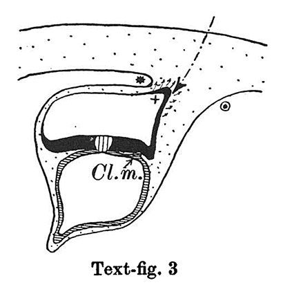
Text-fig 3. Schemes of the chief stages in the development of the human embryo
In stage 3 (text-fig.3, corresponding approximately with the Fetzer embryo (Fetzer-Florian, 1929)) both the amnio-embryonal and the umbilical stalks are quite distinct. The axis of the connecting stalk has now come to liebehind the caudal end of the embryonic plate and it is beginning to form a curve with the convexity directed caudally. The amniotic cavity runs out into a distinct amniotic tip caudally and dorsally, a feature which is to be seen in all following stages. The extension of the amniotic cavity towards the tissue of the connecting stalk takes place in an opposite direction to that of the embryonic plate, the result being, that the distance between the amniotic tip on the one handandthe chorionic ectoderm and the caudal limit of the amnio-embryonal stalk on the other increases at a much slower rate than the distance between the cranial end of the embryonic plate and the above-mentioned structures. In this stage, approximately in the middle of the embryonic plate, there is present a distinct primordium of the primitive streak and, close to the caudal end of the plate,the cloacal membrane is already developed. Itis noteworthy that these two structures are in no way connected in their origin in the human embryo. The primitive streak primordium seems to correspond in its position with Hensen's knot. in this stage,theyolk-sacdoesnotsend any distinct diverticulum towards the stalk.
Ectoderm black; entoderm lined horizontally, primitive streak vertically, head process and chorda plate obliquely. Mesoderm dotted. The axis of the connecting stalk is marked by an arrow. The limits of the connecting stalk are marked by * and ʘ, the limits of the umbilical stalk by + and ʘ). The direction of the extension of the amniotic cavity towards the connecting stalk is marked by arrows. The parts of the allantois and of the axis of the connecting stalk situated out of the median plane are finely dotted. A. Connecting stalk; B. umbilical stalk; Cl.m. cloacal membrane.
| Historic Disclaimer - information about historic embryology pages |
|---|
 |
- Links: Text-fig 1 | Text-fig 2 | Text-fig 3 | Text-fig 4 | Text-fig 5 | Text-fig 6 | Text-fig 7 | Text-fig 1-7 | Text-fig 8 | Text-fig 9 | Text-fig 8-9 | Florian 1930 | Historic Embryology Papers
Reference
Florian J. The formation of the connecting stalk and the extension of the amniotic cavity towards the tissue of the connecting stalk in young human embryos. (1930) J. Anat., 64: 454-476.
Cite this page: Hill, M.A. (2024, April 25) Embryology Florian1930-text-fig03.jpg. Retrieved from https://embryology.med.unsw.edu.au/embryology/index.php/File:Florian1930-text-fig03.jpg
- © Dr Mark Hill 2024, UNSW Embryology ISBN: 978 0 7334 2609 4 - UNSW CRICOS Provider Code No. 00098G
File history
Click on a date/time to view the file as it appeared at that time.
| Date/Time | Thumbnail | Dimensions | User | Comment | |
|---|---|---|---|---|---|
| current | 18:58, 23 September 2015 | | 407 × 425 (25 KB) | Z8600021 (talk | contribs) |
You cannot overwrite this file.
File usage
The following 3 pages use this file: