File:Flint1906 plate01.jpg

Original file (1,280 × 843 pixels, file size: 149 KB, MIME type: image/jpeg)
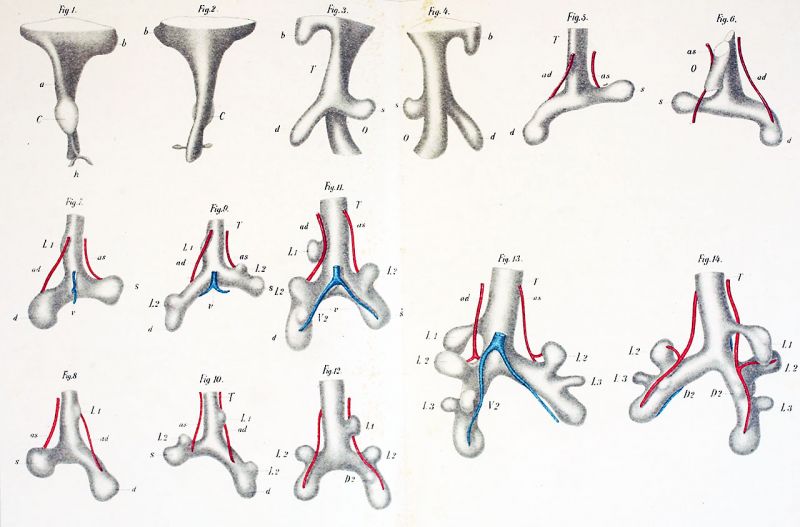
Plate I
Figs. 1-14. are magnified 50 diameters. Pulmonary arteries red, pulmonary veins blue, bronchi white.
Fig. 1. Reconstruction of a portion of the head gut of a pig’s embryo 3 mm. long. Ventral view.
Fig. 2. Dorsal View of the same reconstruction.
Fig. 3. Reconstruction of a. portion of the head gut of a. pig's embryo 5 mm. long. Ventral view.
Fig. 4. Dorsal view of the same reconstruction.
Fig. 5 Reconstruction of the bronchial tree of a pig 6 mm. long.
Fig. 6. Dorsal view of the same reconstruction.
Fig. 7 Reconstruction of the bronchial tree of a. pig 7.5 mm. long.
Fig. 8 Dorsal View of the same reconstruction.
Fig. 9. Reconstruction of the bronchial tree of a. pig 8.5 mm. long.
Fig. 10. Dorsal View of the same reconstruction.
Fig. 11. Reconstruction of the bronchial tree of a pig 10 mm. long.
Fig. 12. Dorsal view of the same reconstruction.
Fig. 13. Reconstruction of the bronchial tree of a pig 12 mm. long.
Fig. 14. Dorsal view of the same reconstruction.
Reference
Flint JM. The development of the lungs. (1906) Amer. J Anat. 6: 1-137.
Cite this page: Hill, M.A. (2024, April 19) Embryology Flint1906 plate01.jpg. Retrieved from https://embryology.med.unsw.edu.au/embryology/index.php/File:Flint1906_plate01.jpg
- © Dr Mark Hill 2024, UNSW Embryology ISBN: 978 0 7334 2609 4 - UNSW CRICOS Provider Code No. 00098G
File history
Click on a date/time to view the file as it appeared at that time.
| Date/Time | Thumbnail | Dimensions | User | Comment | |
|---|---|---|---|---|---|
| current | 14:17, 8 April 2020 | | 1,280 × 843 (149 KB) | Z8600021 (talk | contribs) | adjust size, sharpness |
| 14:16, 8 April 2020 |  | 3,197 × 2,106 (531 KB) | Z8600021 (talk | contribs) | adjust colours | |
| 14:14, 8 April 2020 |  | 3,549 × 2,699 (525 KB) | Z8600021 (talk | contribs) | ==Plate I== figs. 1-14. fiGS. 1-20 are magnified 50 diameters. Pulmonary arteries red, pulmonary veins blue, bronchi white. fiG. 1. Reconstruction of a portion of the head gut of a pig’s embryo 3 mm. long. Ventral view. fiG. 2. Dorsal View of the same reconstruction. fiG. 3. Reconstruction of a. portion of the head gut of a. pig's embryo 5 mm. long. Ventral view. fiG. 4. Dorsal view of the same reconstruction. fiG. 5 Reconstruction of the bronchial tree of a pig 6 mm. long. fiG.... |
You cannot overwrite this file.
File usage
The following page uses this file: