File:Finley1923 Plate 1.jpg
Original file (776 × 1,000 pixels, file size: 151 KB, MIME type: image/jpeg)
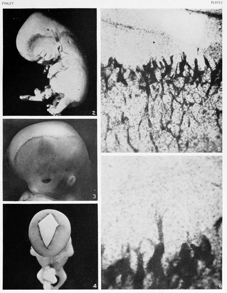
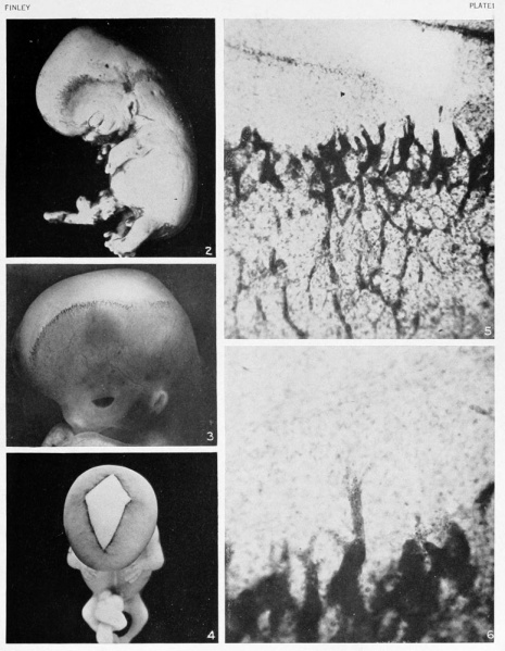
Plate 1
Fig. 2. Photograph of a human embryo 23 mm. in length (No. 966), showing the vascular plexus in the subcutaneous tissue of the head in its earliest form. It is characterized by two distinct growth centers, the temporofrontal and the occipital, from which the vessels gradually spread over the apex of the head. A sharplj defined area between the two growth centers constitutes an angle of retarded growth. X4.
Fig. 3. Photograph of a human embryo 27.5 mm. in length (No. 2561), showing a later stage of the plexus. The angle of retarded growth is not as prominent and the margin of the plexus appears as a narrower and more well-defined line than that in figure 2. X4.
Fig. 4. Photograph of a human embryo 36 mm. in length (No. 1591), showing a late stage in the closing in of the plexus. X2.
Fig. 5. Photograph from a total mount of a piece of the scalp from a human embryo 28 mm. in length (No. 1240a). The varied forms of the growing tips are well shown and the transition from the angioblastic net to the capillary net can easily be followed. X80.
Fig. 6. Photograph from another portion of the same section as above, showing, under higher magnification, the first and second zones. In the center a long tip from the angioblastic plexus is seen to penetrate the avascular zone. This represents the first step in the differentiation of the mesenchyme into angioblastic tissue. X 150.
- 1923 Head Subcutaneous Plexus: Plate 1 | Plate 2 | Fig 1 | Fig 2 | Fig 3 | Fig 4 | Fig 5 | Fig 6 | Fig 7 | Fig 8 | Fig 9 | Fig 10 | Fig 11 | Fig 12 | Fig 13 | Carnegie No.71 | Historic Disclaimer
| Historic Disclaimer - information about historic embryology pages |
|---|
 |
Reference
Finley EB. The development of the subcutaneous vascular plexus in the head of the human embryo. (1923) Contributions to Embryology Carnegie Institution No. 71: 155-161.
Cite this page: Hill, M.A. (2024, April 23) Embryology Finley1923 Plate 1.jpg. Retrieved from https://embryology.med.unsw.edu.au/embryology/index.php/File:Finley1923_Plate_1.jpg
- © Dr Mark Hill 2024, UNSW Embryology ISBN: 978 0 7334 2609 4 - UNSW CRICOS Provider Code No. 00098G
File history
Click on a date/time to view the file as it appeared at that time.
| Date/Time | Thumbnail | Dimensions | User | Comment | |
|---|---|---|---|---|---|
| current | 15:47, 12 August 2012 | | 776 × 1,000 (151 KB) | Z8600021 (talk | contribs) |
You cannot overwrite this file.