File:Fetal pineal gland 01.jpg
Original file (700 × 603 pixels, file size: 67 KB, MIME type: image/jpeg)
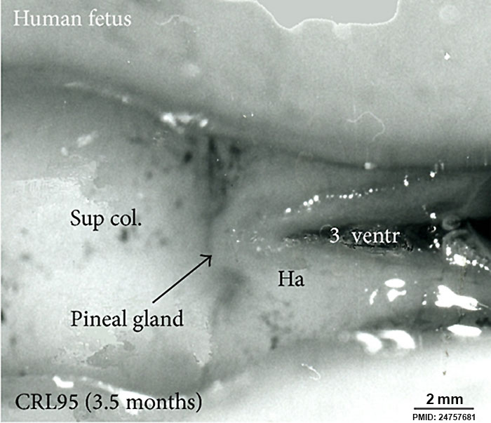
Fetal Pineal Gland Anatomy
Superior (dorsal) view of the diencephalic-mesencephalic area of a 3.5-month-old human fetus.
The third ventricle (3 ventr) without pial covering is seen to the right in the micrograph. The small pineal gland is a small protuberance (arrow) and merging via the broad stalk with the habenula (Ha). Sup col.: superior colliculus. Bar = 2 mm.
Reference
Møller M, Phansuwan-Pujito P & Badiu C. (2014). Neuropeptide Y in the adult and fetal human pineal gland. Biomed Res Int , 2014, 868567. PMID: 24757681 DOI.
Copyright
© 2014 Morten Møller et al. This is an open access article distributed under the Creative Commons Attribution License, which permits unrestricted use, distribution, and reproduction in any medium, provided the original work is properly cited.
Figure 3: 868567.fig.003.jpg http://www.hindawi.com/journals/bmri/2014/868567/fig3 Figure adjusted in size, sharpness and labelling.
Cite this page: Hill, M.A. (2024, April 25) Embryology Fetal pineal gland 01.jpg. Retrieved from https://embryology.med.unsw.edu.au/embryology/index.php/File:Fetal_pineal_gland_01.jpg
- © Dr Mark Hill 2024, UNSW Embryology ISBN: 978 0 7334 2609 4 - UNSW CRICOS Provider Code No. 00098G
File history
Click on a date/time to view the file as it appeared at that time.
| Date/Time | Thumbnail | Dimensions | User | Comment | |
|---|---|---|---|---|---|
| current | 09:05, 13 September 2014 | | 700 × 603 (67 KB) | Z8600021 (talk | contribs) | Superior (dorsal) view of the diencephalic-mesencephalic area of a 3.5-month-old human fetus. The third ventricle (3 ventr) without pial covering is seen to the right in the micrograph. The small pineal gland is a small protuberance (arrow) and mergi... |
You cannot overwrite this file.
File usage
The following 4 pages use this file: